| Citation: | LIU Sicong, LIU Hongzhi, YIN Yaran. Research advances in 3D printed bone tissue engineering scaffolds based on biodegradable polyester/bioceramics[J]. Acta Materiae Compositae Sinica, 2024, 41(4): 1672-1693. doi: 10.13801/j.cnki.fhclxb.20231211.002

|

| [1] |
WANG W, YEUNG K W K. Bone grafts and biomaterials substitutes for bone defect repair: A review[J]. Bioactive Materials, 2017, 2(4): 224-247. doi: 10.1016/j.bioactmat.2017.05.007
|
| [2] |
GIANNOUDIS P V, DINOPOULOS H, TSIRIDIS E. Bone substitutes: An update[J]. Injury, 2005, 36: S20-S27.
|
| [3] |
TAKIZAWA T, NAKAYAMA N, HANIU H, et al. Titanium fiber plates for bone tissue repair[J]. Advanced Materials, 2018, 30(4): 1703608. doi: 10.1002/adma.201703608
|
| [4] |
DIMITRIOU R, MATALIOTAKIS G I, ANGOULES A G, et al. Complications following autologous bone graft harvesting from the iliac crest and using the RIA: A systematic review[J]. Injury, 2011, 42: S3-S15.
|
| [5] |
BERGSMA E J, ROZEMA F R, BOS R R, et al. Foreign body reactions to resorbable poly(L-lactide) bone plates and screws used for the fixation of unstable zygomatic fractures[J]. Journal of Oral and Maxillofacial Surgery, 1993, 51(6): 666-670.
|
| [6] |
SURMENEVA M A, CHAIKINA M V, ZAIKOVSKIY V, et al. The structure of an RF-magnetron sputter-deposited silicate-containing hydroxyapatite-based coating investigated by high-resolution techniques[J]. Surface & Coatings Technology, 2013, 218: 39-46.
|
| [7] |
LEGEROS R Z. Calcium phosphate materials in restorative dentistry: A review[J]. Advances in Dental Research, 1988, 2(1): 164-180. doi: 10.1177/08959374880020011101
|
| [8] |
LEBEDEV S M, GEFLE O S, AMITOV E T, et al. Mechanical properties of PLA-based composites for fused deposition modeling technology[J]. The International Journal of Advanced Manufacturing Technology, 2018, 97(1): 511-518.
|
| [9] |
REN X, LIU Q, ZHENG S, et al. Synergistic delivery of bFGF and BMP-2 from poly(L-lactic-co-glycolic acid)/graphene oxide/hydroxyapatite nanofibre scaffolds for bone tissue engineering applications[J]. RSC Advances, 2018, 8(56): 31911-31923. doi: 10.1039/C8RA05250F
|
| [10] |
COSTANTINI M, BARBETTA A. 6-gas foaming technologies for 3D scaffold engineering[M]//DENG Y, KUIPER J. Functional 3D Tissue Engineering Scaffolds. Cambridge: Woodhead Publishing, 2018: 127-149.
|
| [11] |
COOPER A I. Polymer synthesis and processing using supercritical carbon dioxide[J]. Journal of Materials Chemistry, 2000, 10(2): 207-234. doi: 10.1039/a906486i
|
| [12] |
PANKONGADISAK P, JAIKAEW N, KITI K, et al. The potential use of gentamicin sulfate-loaded poly(L-lactic acid)-sericin hybrid scaffolds for bone tissue engineering[J]. Polymer Bulletin, 2019, 76(6): 2867-2885. doi: 10.1007/s00289-018-2520-x
|
| [13] |
BUZAROVSKA A, DINESCU S, CHITOIU L, et al. Porous poly(L-lactic acid) nanocomposite scaffolds with functionalized TiO2 nanoparticles: Properties, cytocompatibility and drug release capability[J]. Journal of Materials Science, 2018, 53(16): 11151-11166. doi: 10.1007/s10853-018-2415-0
|
| [14] |
PUPPI D, CHIELLINI F, PIRAS A M, et al. Polymeric materials for bone and cartilage repair[J]. Progress in Polymer Science, 2010, 35(4): 403-440. doi: 10.1016/j.progpolymsci.2010.01.006
|
| [15] |
ZHANG X, CHEN J L, XING F, et al. Three-dimensional printed polylactic acid and hydroxyapatite composite scaffold with urine-derived stem cells as a treatment for bone defects[J]. Journal of Materials Science: Materials in Medicine, 2022, 33(10): 71. doi: 10.1007/s10856-022-06686-z
|
| [16] |
SENATOV F S, NIAZA K V, ZADOROZHNYY M Y, et al. Mechanical properties and shape memory effect of 3D-printed PLA-based porous scaffolds[J]. Journal of the Mechanical Behavior of Biomedical Materials, 2016, 57: 139-148. doi: 10.1016/j.jmbbm.2015.11.036
|
| [17] |
HEIDARI-RARANI M, RAFIEE-AFARANI M, ZAHEDI A M. Mechanical characterization of FDM 3D printing of continuous carbon fiber reinforced PLA composites[J]. Composites Part B: Engineering, 2019, 175: 107147. doi: 10.1016/j.compositesb.2019.107147
|
| [18] |
CHINNASAMI H, DEY M K, DEVIREDDY R. Three-dimensional scaffolds for bone tissue engineering[J]. Bioengineering (Basel), 2023, 10(7): 759. doi: 10.3390/bioengineering10070759
|
| [19] |
SHUAI C, YANG F, SHUAI Y, et al. Silicon dioxide nanoparticles decorated on graphene oxide nanosheets and their application in poly(L-lactic acid) scaffold[J]. Journal of Advanced Research, 2023, 48: 175-190. doi: 10.1016/j.jare.2022.08.017
|
| [20] |
ZHOU X, ZHOU G, JUNKA R, et al. Fabrication of polylactic acid (PLA)-based porous scaffold through the combination of traditional bio-fabrication and 3D printing technology for bone regeneration[J]. Colloids and Surfaces B: Biointerfaces, 2021, 197: 111420. doi: 10.1016/j.colsurfb.2020.111420
|
| [21] |
ZHAO D, WANG Y, YU Z, et al. Co-culture bioprinting of tissue-engineered bone-periosteum biphasic complex for repairing critical-sized skull defects in rabbits[J]. International Journal of Bioprinting, 2023, 9: 698. doi: 10.18063/ijb.698
|
| [22] |
KOONS G L, DIBA M, MIKOS A G. Materials design for bone-tissue engineering[J]. Nature Reviews Materials, 2020, 5(8): 584-603. doi: 10.1038/s41578-020-0204-2
|
| [23] |
TRIVEDI A K, GUPTA M K, SINGH H. PLA based biocomposites for sustainable products: A review[J]. Advanced Industrial and Engineering Polymer Research, 2023, 6(4): 382-395. doi: 10.1016/j.aiepr.2023.02.002
|
| [24] |
SMIESZEK A, TOMASZEWSKI K A, KORNICKA K, et al. Metformin promotes osteogenic differentiation of adipose-derived stromal cells and exerts pro-osteogenic effect stimulating bone regeneration[J]. Journal of Clinical Medicine, 2018, 7(12): 482. doi: 10.3390/jcm7120482
|
| [25] |
CHEN X, FAN H, DENG X, et al. Scaffold structural microenvironmental cues to guide tissue regeneration in bone tissue applications[J]. Nanomaterials (Basel), 2018, 8(11): 960. doi: 10.3390/nano8110960
|
| [26] |
WUBNEH A, TSEKOURA E K, AYRANCI C, et al. Current state of fabrication technologies and materials for bone tissue engineering[J]. Acta Biomaterialia, 2018, 80: 1-30. doi: 10.1016/j.actbio.2018.09.031
|
| [27] |
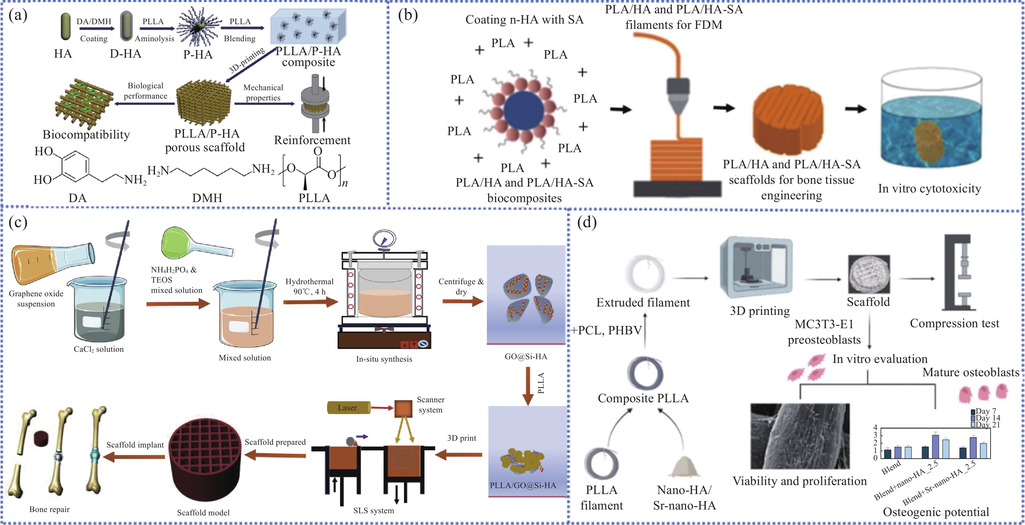
YANG W F, LONG L, WANG R, et al. Surface-modified hydroxyapatite nanoparticle-reinforced polylactides for three-dimensional printed bone tissue engineering scaffolds[J]. Journal of Biomedical Nanotechnology, 2018, 14(2): 294-303. doi: 10.1166/jbn.2018.2495
|
| [28] |
ZHANG B, WANG L, SONG P, et al. 3D printed bone tissue regenerative PLA/HA scaffolds with comprehensive performance optimizations[J]. Materials & Design, 2021, 201: 109490.
|
| [29] |
CÉ DE ANDRADE J, CABRAL F, CLEMENS F J, et al. Effect of stearic acid on the mechanical and rheological properties of PLA/HA biocomposites[J]. Materials Today Communications, 2023, 35: 106357. doi: 10.1016/j.mtcomm.2023.106357
|
| [30] |
SONG X, GUAN W, QIN H, et al. Properties of poly(lactic acid)/walnut shell/hydroxyapatite composites prepared with fused deposition modeling[J]. Scientific Reports, 2022, 12(1): 11563. doi: 10.1038/s41598-022-15622-8
|
| [31] |
WANG G, QIAN G, ZAN J, et al. A co-dispersion nanosystem of graphene oxide@silicon-doped hydroxyapatite to improve scaffold properties[J]. Materials & Design, 2021, 199: 109399.
|
| [32] |
KONTOGIANNI G I, BONATTI A F, DE MARIA C, et al. Promotion of in vitro osteogenic activity by melt extrusion-based PLLA/PCL/PHBV scaffolds enriched with nano-hydroxyapatite and strontium substituted nano-hydroxyapatite[J]. Polymers (Basel), 2023, 15(4): 1052. doi: 10.3390/polym15041052
|
| [33] |
SAHMANI S, KHANDAN A, SABER-SAMANDARI S, et al. Fabrication and resonance simulation of 3D-printed biocomposite mesoporous implants with different periodic cellular topologies[J]. Bioprinting, 2021, 22: e00138. doi: 10.1016/j.bprint.2021.e00138
|
| [34] |
PREMPHET P, LEKSAKUL K, BOONYAWAN D, et al. Process parameters optimization and mechanical properties of 3D PLA/HA printing scaffold[J]. Materials Today: Proceedings, 2023, 4: 124.
|
| [35] |
HWANGBO H, LEE J, KIM G. Mechanically and biologically enhanced 3D-printed HA/PLLA/dECM biocomposites for bone tissue engineering[J]. International Journal of Biological Macromolecules, 2022, 218: 9-21. doi: 10.1016/j.ijbiomac.2022.07.040
|
| [36] |
PÉREZ-DAVILA S, GARRIDO-GULÍAS N, GONZÁLEZ-RODRÍGUEZ L, et al. Physicochemical properties of 3D-printed polylactic acid/hydroxyapatite scaffolds[J]. Polymers (Basel), 2023, 15(13): 2849. doi: 10.3390/polym15132849
|
| [37] |
FU Z, CUI J, ZHAO B, et al. An overview of polyester/hydroxyapatite composites for bone tissue repairing[J]. Journal of Orthopaedic Translation, 2021, 28: 118-130. doi: 10.1016/j.jot.2021.02.005
|
| [38] |
BACKES E H, FERNANDES E M, DIOGO G S, et al. Engineering 3D printed bioactive composite scaffolds based on the combination of aliphatic polyester and calcium phosphates for bone tissue regeneration[J]. Materials Science and Engineering: C, 2021, 122: 111928. doi: 10.1016/j.msec.2021.111928
|
| [39] |
SHUAI C J, YANG W J, FENG P, et al. Accelerated degradation of HAP/PLLA bone scaffold by PGA blending facilitates bioactivity and osteoconductivity[J]. Bioactive Materials, 2021, 6(2): 490-502. doi: 10.1016/j.bioactmat.2020.09.001
|
| [40] |
FENG P, PENG S, SHUAI C, et al. In situ generation of hydroxyapatite on biopolymer particles for fabrication of bone scaffolds owning bioactivity[J]. ACS Applied Materials & Interfaces, 2020, 12(41): 46743-46755.
|
| [41] |
BAHRAMINASAB M, DOOSTMOHAMMADI N, TALEBI A, et al. 3D printed polylactic acid/gelatin-nano-hydroxyapatite/platelet-rich plasma scaffold for critical-sized skull defect regeneration[J]. BioMedical Engineering OnLine, 2022, 21(1): 86. doi: 10.1186/s12938-022-01056-w
|
| [42] |
NARAYANAN G, VERNEKAR V N, KUYINU E L, et al. Poly (lactic acid)-based biomaterials for orthopaedic regenerative engineering[J]. Advanced Drug Delivery Reviews, 2016, 107: 247-276. doi: 10.1016/j.addr.2016.04.015
|
| [43] |
CHIA H N, WU B M. Recent advances in 3D printing of biomaterials[J]. Journal of Biological Engineering, 2015, 9: 4. doi: 10.1186/s13036-015-0001-4
|
| [44] |
HARRIS J S, BEMENDERFER T B, WESSEL A R, et al. A review of mouse critical size defect models in weight bearing bones[J]. Bone, 2013, 55(1): 241-247. doi: 10.1016/j.bone.2013.02.002
|
| [45] |
WEI L, WU S, KUSS M, et al. 3D printing of silk fibroin-based hybrid scaffold treated with platelet rich plasma for bone tissue engineering[J]. Bioactive Materials, 2019, 4: 256-260. doi: 10.1016/j.bioactmat.2019.09.001
|
| [46] |
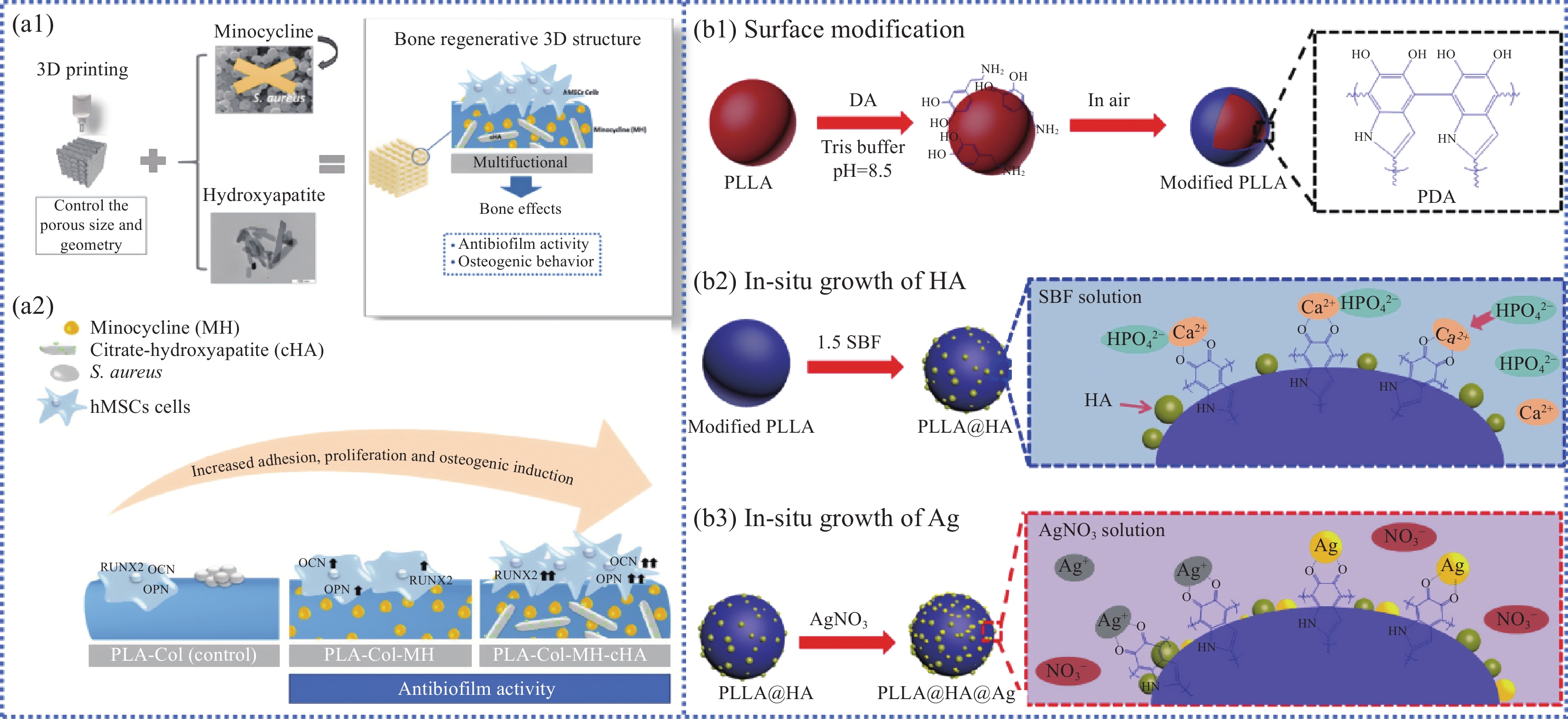
MARTIN V, RIBEIRO I A, ALVES M M, et al. Engineering a multifunctional 3D-printed PLA-collagen-minocycline-nanohydroxyapatite scaffold with combined antimicrobial and osteogenic effects for bone regeneration[J]. Materials Science and Engineering: C, 2019, 101: 15-26. doi: 10.1016/j.msec.2019.03.056
|
| [47] |
YANG Y, CHENG Y, DENG F, et al. A bifunctional bone scaffold combines osteogenesis and antibacterial activity via in situ grown hydroxyapatite and silver nanoparticles[J]. Bio-Design and Manufacturing, 2021, 4(3): 452-468. doi: 10.1007/s42242-021-00130-x
|
| [48] |
MARYCZ K, SMIESZEK A, TARGONSKA S, et al. Three dimensional (3D) printed polylactic acid with nano-hydroxyapatite doped with europium(III) ions (nHAp/PLLA@Eu3+) composite for osteochondral defect regeneration and theranostics[J]. Materials Science and Engineering: C, 2020, 110: 110634. doi: 10.1016/j.msec.2020.110634
|
| [49] |
ZHENG H, SUN Z, ZHANG H. Effects of walnut shell powders on the morphology and the thermal and mechanical properties of poly(lactic acid)[J]. Journal of Thermoplastic Composite Materials, 2019, 33(10): 1383-1395.
|
| [50] |
FERREIRA F V, BRITO F S, FRANCESCHI W, et al. Functionalized graphene oxide as reinforcement in epoxy based nanocomposites[J]. Surfaces and Interfaces, 2018, 10: 100-109. doi: 10.1016/j.surfin.2017.12.004
|
| [51] |
VILA M, GARCÍA A, GIROTTI A, et al. 3D silicon doped hydroxyapatite scaffolds decorated with elastin-like recombinamers for bone regenerative medicine[J]. Acta Biomaterialia, 2016, 45: 349-356. doi: 10.1016/j.actbio.2016.09.016
|
| [52] |
HOU Y, WANG W, BARTOLO P. Investigation of polycaprolactone for bone tissue engineering scaffolds: In vitro degradation and biological studies[J]. Materials & Design, 2022, 216: 110582.
|
| [53] |
JIAO Z, LUO B, XIANG S, et al. 3D printing of HA/PCL composite tissue engineering scaffolds[J]. Advanced Industrial and Engineering Polymer Research, 2019, 2(4): 196-202. doi: 10.1016/j.aiepr.2019.09.003
|
| [54] |
JOHN J, DEVJANI D, ALI S, et al. Optimization of 3D printed polylactic acid structures with different infill patterns using Taguchi-grey relational analysis[J]. Advanced Industrial and Engineering Polymer Research, 2023, 6(1): 62-78. doi: 10.1016/j.aiepr.2022.06.002
|
| [55] |
ESHRAGHI S, DAS S. Micromechanical finite-element modeling and experimental characterization of the compressive mechanical properties of polycaprolactone-hydroxyapatite composite scaffolds prepared by selective laser sintering for bone tissue engineering[J]. Acta Biomaterialia, 2012, 8(8): 3138-3143. doi: 10.1016/j.actbio.2012.04.022
|
| [56] |
OSTAFINSKA A, FORTELNÝ I, HODAN J, et al. Strong synergistic effects in PLA/PCL blends: Impact of PLA matrix viscosity[J]. Journal of the Mechanical Behavior of Biomedical Materials, 2017, 69: 229-241. doi: 10.1016/j.jmbbm.2017.01.015
|
| [57] |
OLEWNIK-KRUSZKOWSKA E, KASPERSKA P, KOTER I. Effect of poly(ε-caprolactone) as plasticizer on the properties of composites based on polylactide during hydrolytic degradation[J]. Reactive and Functional Polymers, 2016, 103: 99-107. doi: 10.1016/j.reactfunctpolym.2016.03.026
|
| [58] |
ÅKERLUND E, DIEZ-ESCUDERO A, GRZESZCZAK A, et al. The effect of PCL addition on 3D-printable PLA/HA composite filaments for the treatment of bone defects[J]. Polymers (Basel), 2022, 14(16): 3305. doi: 10.3390/polym14163305
|
| [59] |
ESPOSITO CORCIONE C, GERVASO F, SCALERA F, et al. Highly loaded hydroxyapatite microsphere/PLA porous scaffolds obtained by fused deposition modelling[J]. Ceramics International, 2019, 45(2): 2803-2810.
|
| [60] |
JOSEPH B, NINAN N, VISALAKSHAN R M, et al. Insights into the biomechanical properties of plasma treated 3D printed PCL scaffolds decorated with gold nanoparticles[J]. Composites Science and Technology, 2021, 202: 108544. doi: 10.1016/j.compscitech.2020.108544
|
| [61] |
LIU C, YAO W, TIAN M, et al. Mussel-inspired degradable antibacterial polydopamine/silica nanoparticle for rapid hemostasis[J]. Biomaterials, 2018, 179: 83-95. doi: 10.1016/j.biomaterials.2018.06.037
|
| [62] |
WANG H, YANG J, LIU X, et al. A robust 3D superhydrophobic sponge for in situ continuous oil removing[J]. Journal of Materials Science, 2019, 54(2): 1255-1266. doi: 10.1007/s10853-018-2938-4
|
| [63] |
DOU Y, HUANG J, XIA X, et al. A hierarchical scaffold with a highly pore-interconnective 3D printed PLGA/n-HA framework and an extracellular matrix like gelatin network filler for bone regeneration[J]. Journal of Materials Chemistry B, 2021, 9(22): 4488-4501. doi: 10.1039/D1TB00662B
|
| [64] |
DAVIDENKO N, SCHUSTER C F, BAX D V, et al. Evaluation of cell binding to collagen and gelatin: A study of the effect of 2D and 3D architecture and surface chemistry[J]. Journal of Materials Science-Materials in Medicine, 2016, 27(10): 148. doi: 10.1007/s10856-016-5763-9
|
| [65] |
CHENG W X, LIU Y Z, MENG X B, et al. PLGA/β-TCP composite scaffold incorporating cucurbitacin B promotes bone regeneration by inducing angiogenesis[J]. Journal of Orthopaedic Translation, 2021, 31: 41-51. doi: 10.1016/j.jot.2021.10.002
|
| [66] |
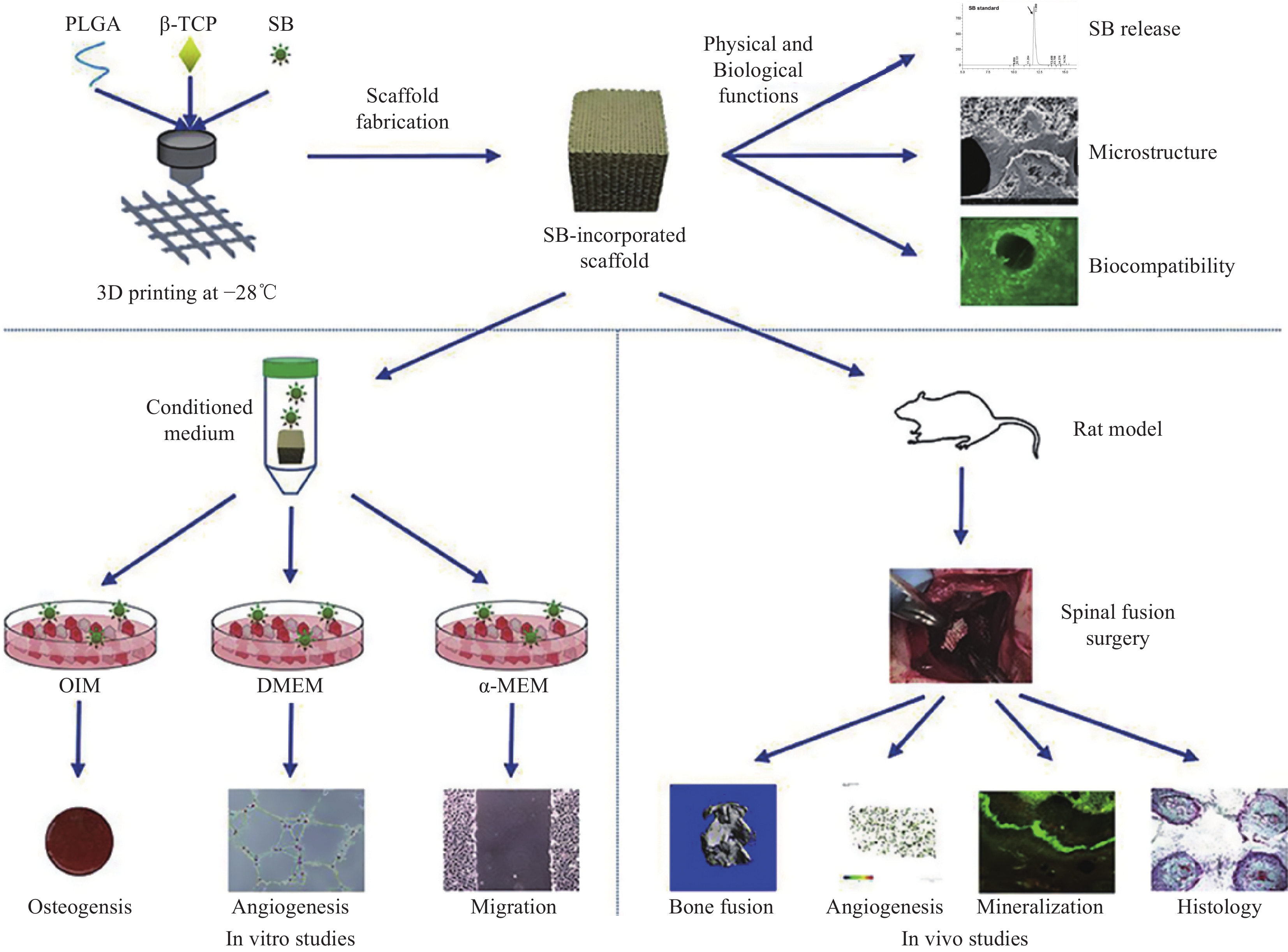
LIN S, CUI L, CHEN G, et al. PLGA/β-TCP composite scaffold incorporating salvianolic acid B promotes bone fusion by angiogenesis and osteogenesis in a rat spinal fusion model[J]. Biomaterials, 2019, 196: 109-121. doi: 10.1016/j.biomaterials.2018.04.004
|
| [67] |
ZHU W, LIANG S, WANG J, et al. Europium-phenolic network coated BaGdF5 nanocomposites for tri-modal computed tomography/magnetic resonance/luminescence imaging[J]. Journal of Materials Science: Materials in Medicine, 2017, 28(5): 74. doi: 10.1007/s10856-017-5888-5
|
| [68] |
HE J, ZHANG N, ZHANG J, et al. Migration critically meditates osteoblastic differentiation of bone mesenchymal stem cells through activating canonical Wnt signal pathway[J]. Colloids and Surfaces B: Biointerfaces, 2018, 171: 205-213. doi: 10.1016/j.colsurfb.2018.07.017
|
| [69] |
WANG W, WEI J, LEI D, et al. 3D printing of lithium osteogenic bioactive composite scaffold for enhanced bone regeneration[J]. Composites Part B: Engineering, 2023, 256: 110641. doi: 10.1016/j.compositesb.2023.110641
|
| [70] |
XU Z, LIN B, ZHAO C, et al. Lanthanum doped octacalcium phosphate/polylactic acid scaffold fabricated by 3D printing for bone tissue engineering[J]. Journal of Materials Science & Technology, 2022, 118: 229-242.
|
| [71] |
HUANG J, LIU W, LIANG Y, et al. Preparation and biocompatibility of diphasic magnetic nanocomposite scaffold[J]. Materials Science and Engineering: C, 2018, 87: 70-77. doi: 10.1016/j.msec.2018.02.003
|
| [72] |
PORTER R M, LIU F, PILAPIL C, et al. Osteogenic potential of reamer irrigator aspirator (RIA) aspirate collected from patients undergoing hip arthroplasty[J]. Journal of Orthopaedic Research, 2009, 27(1): 42-49. doi: 10.1002/jor.20715
|
| [73] |
LIU Z, CHU W, ZHANG L, et al. The effect of enhanced bone marrow in conjunction with 3D-printed PLA-HA in the repair of critical-sized bone defects in a rabbit model[J]. Annals of Translational Medicine, 2021, 9(14): 1134. doi: 10.21037/atm-20-8198
|
| [74] |
PADUANO F, MARRELLI M, ALOM N, et al. Decellularized bone extracellular matrix and human dental pulp stem cells as a construct for bone regeneration[J]. Journal of Biomaterials Science, Polymer Edition, 2017, 28(8): 730-748. doi: 10.1080/09205063.2017.1301770
|
| [75] |
GENDVILIENE I, SIMOLIUNAS E, ALKSNE M, et al. Effect of extracellular matrix and dental pulp stem cells on bone regeneration with 3D printed PLA/HA composite scaffolds[J]. European Cells and Materials, 2021, 41: 204-215. doi: 10.22203/eCM.v041a15
|
| [76] |
ZHANG D, WEI G, LI P, et al. Urine-derived stem cells: A novel and versatile progenitor source for cell-based therapy and regenerative medicine[J]. Genes & Diseases, 2014, 1(1): 8-17.
|
| [77] |
BODIN A, BHARADWAJ S, WU S, et al. Tissue-engineered conduit using urine-derived stem cells seeded bacterial cellulose polymer in urinary reconstruction and diversion[J]. Biomaterials, 2010, 31(34): 8889-8901. doi: 10.1016/j.biomaterials.2010.07.108
|
| [78] |
LIAO H T, LEE M Y, TSAI W W, et al. Osteogenesis of adipose-derived stem cells on polycaprolactone-β-tricalcium phosphate scaffold fabricated via selective laser sintering and surface coating with collagen type I[J]. Journal of Tissue Engineering and Regenerative Medicine, 2016, 10(10): E337-E353. doi: 10.1002/term.1811
|
| [79] |
ZHENG C, ATTARILAR S, LI K, et al. 3D-printed HA15-loaded β-tricalcium phosphate/poly (lactic-co-glycolic acid) bone tissue scaffold promotes bone regeneration in rabbit radial defects[J]. International Journal of Bioprinting, 2021, 7(1): 317.
|
| [80] |
SONG M, ZHAO D, WEI S, et al. The effect of electromagnetic fields on the proliferation and the osteogenic or adipogenic differentiation of mesenchymal stem cells modulated by dexamethasone[J]. Bioelectromagnetics, 2014, 35(7): 479-490. doi: 10.1002/bem.21867
|
| [81] |
TU C, CHEN J, HUANG C, et al. Effects of electromagnetic fields treatment on rat critical-sized calvarial defects with a 3D-printed composite scaffold[J]. Stem Cell Research and Therapy, 2020, 11(1): 433. doi: 10.1186/s13287-020-01954-7
|
| [82] |
ZHANG H, MAO X, ZHAO D, et al. Three dimensional printed polylactic acid-hydroxyapatite composite scaffolds for prefabricating vascularized tissue engineered bone: An in vivo bioreactor model[J]. Scientific Reports, 2017, 7(1): 15255. doi: 10.1038/s41598-017-14923-7
|
| [83] |
LIU Y, MÖLLER B, WILTFANG J, et al. Tissue engineering of a vascularized bone graft of critical size with an osteogenic and angiogenic factor-based in vivo bioreactor[J]. Tissue Engineering Part A, 2014, 20(23-24): 3189-3197. doi: 10.1089/ten.tea.2013.0653
|
| [84] |
MAIA-PINTO M O C, BROCHADO A C B, TEIXEIRA B N, et al. Biomimetic mineralization on 3D printed PLA scaffolds: On the response of human primary osteoblasts spheroids and in vivo implantation[J]. Polymers, 2021, 13(1): 74.
|
| [85] |
KWON D Y, PARK J H, JANG S H, et al. Bone regeneration by means of a three-dimensional printed scaffold in a rat cranial defect[J]. Journal of Tissue Engineering and Regenerative Medicine, 2018, 12(2): 516-528. doi: 10.1002/term.2532
|
| [86] |
TCACENCU I, RODRIGUES N, ALHARBI N, et al. Osseointegration of porous apatite-wollastonite and poly(lactic acid) composite structures created using 3D printing techniques[J]. Materials Science and Engineering: C, 2018, 90: 1-7. doi: 10.1016/j.msec.2018.04.022
|
| [87] |
ZHANG H, MAO X, DU Z, et al. Three dimensional printed macroporous polylactic acid/hydroxyapatite composite scaffolds for promoting bone formation in a critical-size rat calvarial defect model[J]. Science and Technology of Advanced Materials, 2016, 17(1): 136-148. doi: 10.1080/14686996.2016.1145532
|
| [88] |
WANG W, ZHANG B, LI M, et al. 3D printing of PLA/n-HA composite scaffolds with customized mechanical properties and biological functions for bone tissue engineering[J]. Composites Part B: Engineering, 2021, 224: 109192. doi: 10.1016/j.compositesb.2021.109192
|
| [89] |
WANG W, ZHANG B, ZHAO L, et al. Fabrication and properties of PLA/nano-HA composite scaffolds with balanced mechanical properties and biological functions for bone tissue engineering application[J]. Nanotechnology Reviews, 2021, 10(1): 1359-1373. doi: 10.1515/ntrev-2021-0083
|
| [90] |
LIU Z, GE Y, ZHANG L, et al. The effect of induced membranes combined with enhanced bone marrow and 3D PLA-HA on repairing long bone defects in vivo[J]. Journal of Tissue Engineering and Regenerative Medicine, 2020, 14(10): 1403-1414. doi: 10.1002/term.3106
|
| [91] |
CHEN X, GAO C, JIANG J, et al. 3D printed porous PLA/nHA composite scaffolds with enhanced osteogenesis and osteoconductivity in vivo for bone regeneration[J]. Biomedical Materials, 2019, 14(6): 065003. doi: 10.1088/1748-605X/ab388d
|
| [92] |
FERREIRA M, RZHEPISHEVSKA O, GRENHO L, et al. Levofloxacin-loaded bone cement delivery system: Highly effective against intracellular bacteria and Staphylococcus aureus biofilms[J]. International Journal of Pharmaceutics, 2017, 532(1): 241-248. doi: 10.1016/j.ijpharm.2017.08.089
|
| [93] |
SILVA T, GRENHO L, BARROS J, et al. A minocycline-releasing PMMA system as a space maintainer for staged bone reconstructions-in vitro antibacterial, cytocompatibility and anti-inflammatory characterization[J]. Biomedical Materials, 2017, 12(3): 035009. doi: 10.1088/1748-605X/aa68b8
|
| [94] |
LI X, WANG Y, WANG Z, et al. Composite PLA/PEG/nHA/dexamethasone scaffold prepared by 3D printing for bone regeneration[J]. Macromolecular Bioscience, 2018, 18(6): e1800068. doi: 10.1002/mabi.201800068
|
| [95] |
WANG Y, YAN L, CHENG R, et al. Multifunctional HA/Cu nano-coatings on titanium using PPy coordination and doping via pulse electrochemical polymerization[J]. Biomaterials Science, 2018, 6(3): 575-585. doi: 10.1039/C7BM01104K
|
| [96] |
RÊGO D F, ELIAS S T, AMATO A A, et al. Anti-tumor effects of metformin on head and neck carcinoma cell lines: A systematic review[J]. Oncology Letters, 2017, 13(2): 554-566. doi: 10.3892/ol.2016.5526
|
| [97] |
TAN W, GAO C, FENG P, et al. Dual-functional scaffolds of poly(L-lactic acid)/nanohydroxyapatite encapsulated with metformin: Simultaneous enhancement of bone repair and bone tumor inhibition[J]. Materials Science and Engineering: C, 2021, 120: 111592. doi: 10.1016/j.msec.2020.111592
|
| [98] |
SHI M, XIA L, CHEN Z, et al. Europium-doped mesoporous silica nanosphere as an immune-modulating osteogenesis/angiogenesis agent[J]. Biomaterials, 2017, 144: 176-187. doi: 10.1016/j.biomaterials.2017.08.027
|
Figures(8) / Tables(8)


 DownLoad: