


Fabrication of polycaprolactone-cellulose acetate-poly(L-lactic acid) three-dimensional micro-nanofibrous porous scaffold composites and its bio-mineralization activity
-
摘要: 3D纳米纤维多孔支架作为骨组织工程支架材料具有很好的发展前景。在无其它任何添加剂条件下,通过低温相分离方法制备了左旋聚乳酸-聚己内酯-醋酸纤维素(PCL-CA-PLLA)三维微-纳米复合纤维多孔支架材料。采用SEM分析聚合物比例、淬火时间、聚合物浓度和淬火温度等条件对纤维支架材料形貌影响。PCL-CA-PLLA(1∶1∶8)的直径为(276±121) nm,该直径与细胞外基质的尺寸大小(50~500 nm)相当,孔隙率和比表面积分别为95.12%和54.18 m2/g。说明PCL-CA-PLLA三维微-纳米复合纤维多孔支架材料为高孔隙率和大比表面积的三维多孔材料。与纯PLLA纤维支架材料相比,PCL-CA-PLLA三维微-纳米复合纤维多孔支架材料的机械强度有所提高,亲水性有所改善。PCL-CA-PLLA三维微-纳米复合纤维有望成为理想的组织工程支架材料。Abstract: 3D nanofiber scaffold composites in bone tissue engineering are promising. Poly(L-lactic acid)-polycaprolactone-cellulose acetate 3D composite micro-nanofibrous porous scaffolds were prepared by low-temperature phase separation, without the assistance of additives. The effects of PCL-CA-PLLA ratio, quenching time, polymer concentration and quenching temperature on the morphology of fibrous scaffolds were investigated by SEM. The diameter of PCL-CA-PLLA (1∶1∶8) is (276±121) nm, which is similar to the size of the extracellular matrix (50-500 nm), and the porosity and specific surface area are 95.12% and 54.18 m2/g, respectively. It is indicated that PCL-CA-PLLA 3D micro-nanofibrous porous scaffold composites are 3D porous materials with high porosity and large specific surface area. Compared with pure PLLA fibrous scaffolds, the mechanical strength and hydrophilicity of PCL-CA-PLLA 3D micro-nanofibrous porous scaffold composites are improved. PLLA-PCL-CA 3D micro-nanofibrous are expected to be ideal tissue engineering scaffold materials.
-
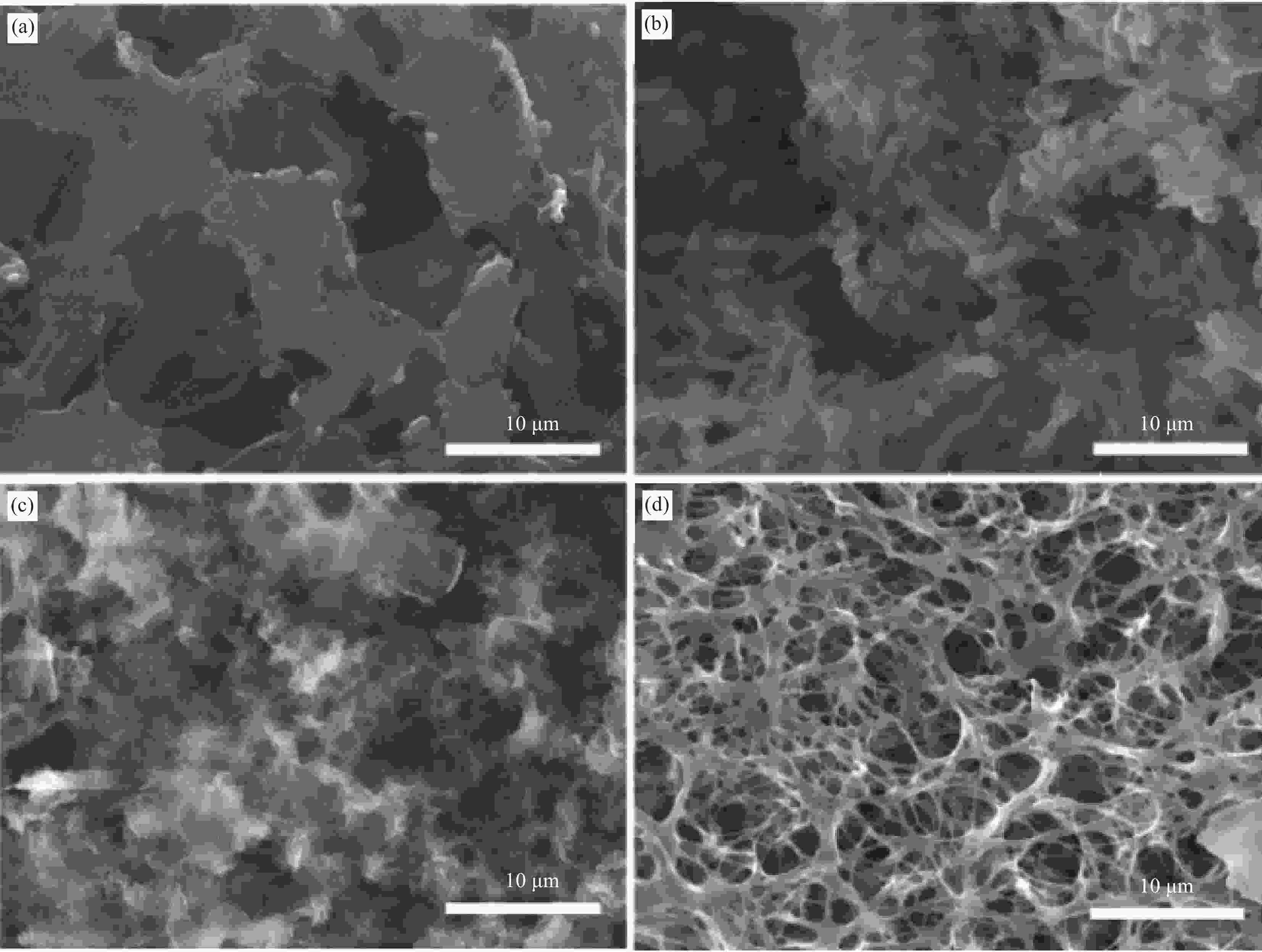
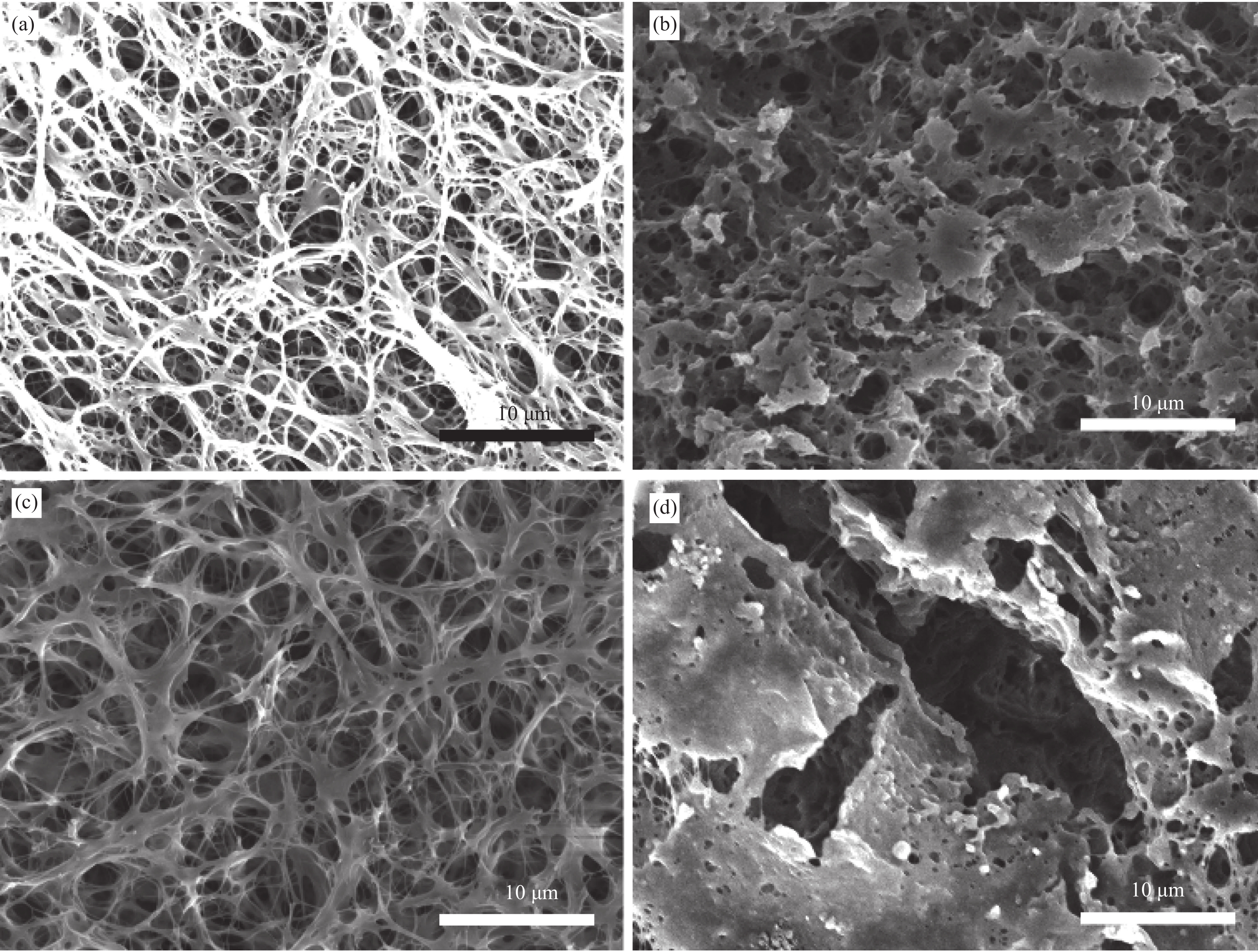
图 1 不同溶剂的PCL-CA-PLLA复合材料的SEM图像
Figure 1. SEM images of PCL-CA-PLLA composites with diffierent solvents((a) DO; (b) Acetonitrile; (c) DMAc; (d) THF)
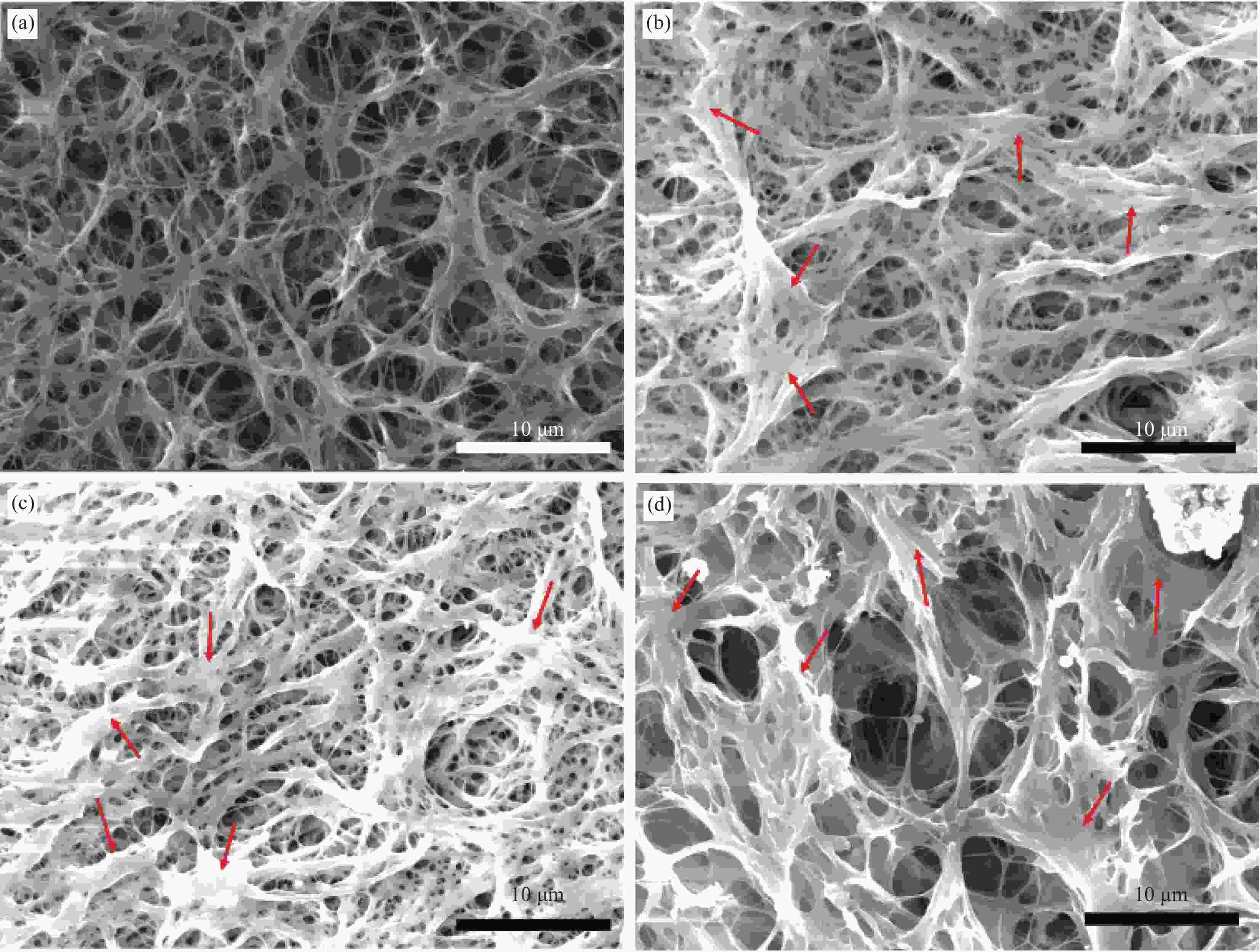
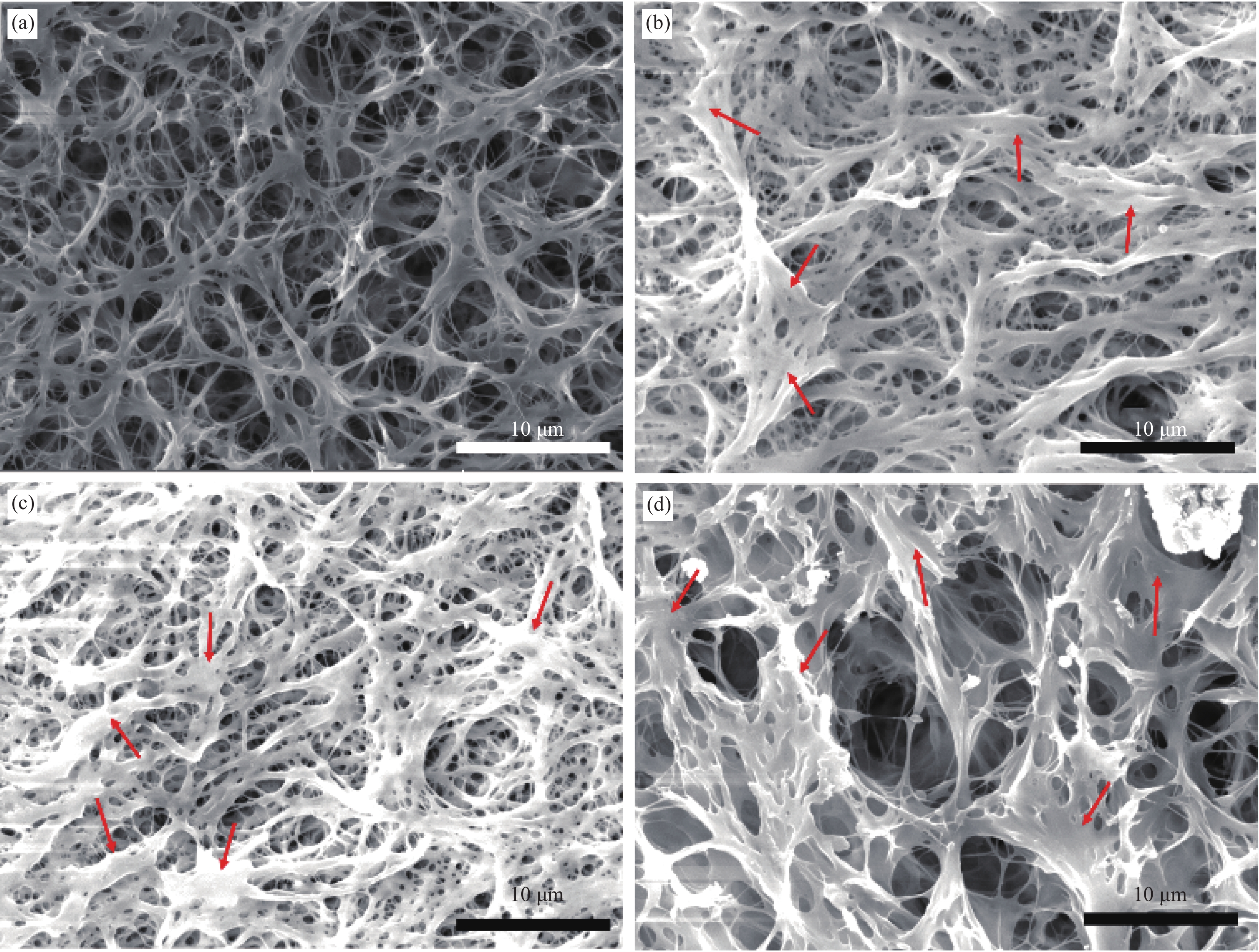
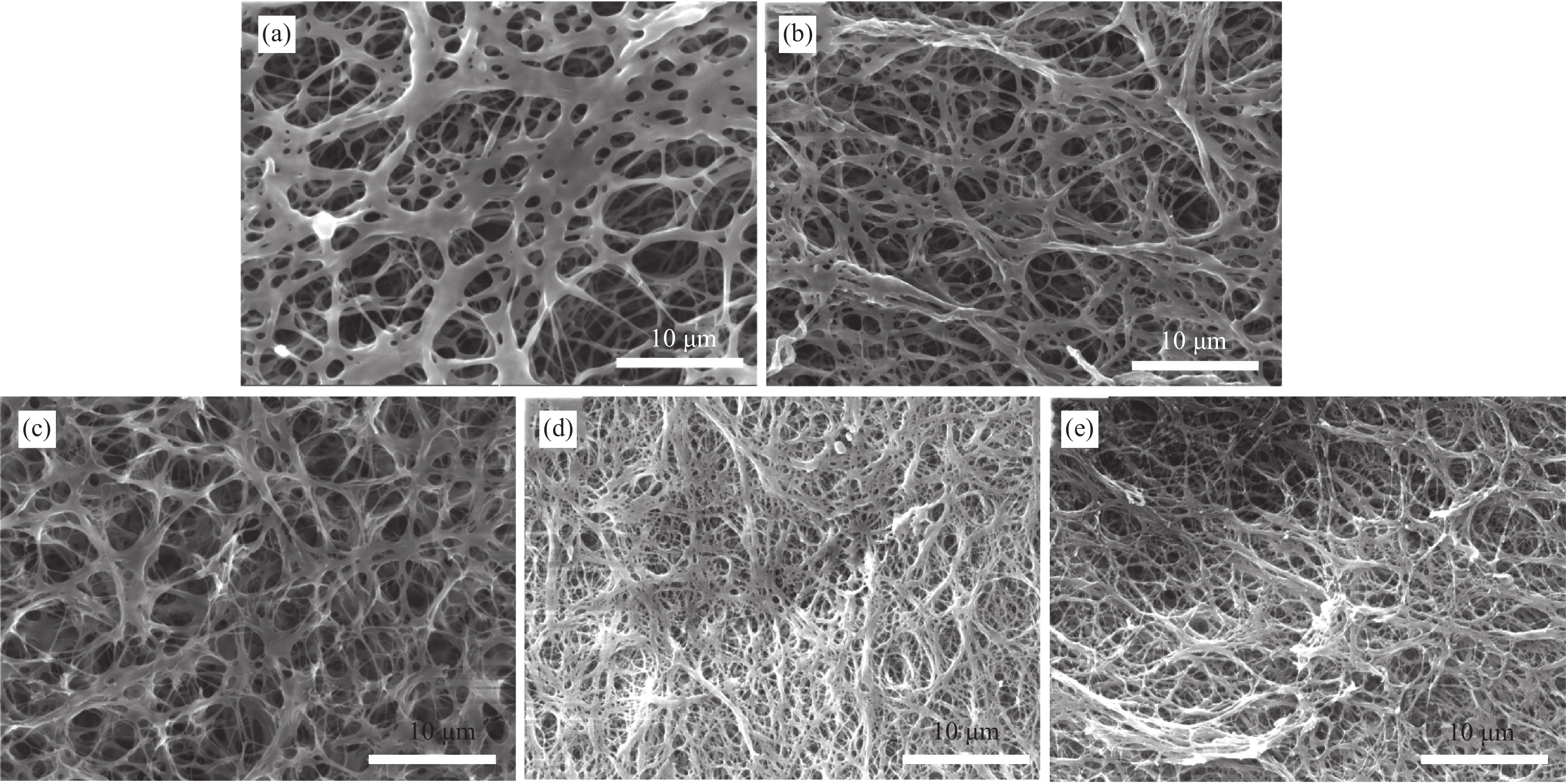
图 2 不同质量比的PCL-CA-PLLA三维微-纳米复合纤维扫描电镜图(其余条件参见表1)
Figure 2. SEM images of PCL-CA-PLLA 3D composites micro-nanofibrous with different mass ratio of PCL-CA-PLLA, (a) 1:1:8,(b) 1.5:1.5:7.5,(c) 2:2:6,(d) 2.5:2.5:6
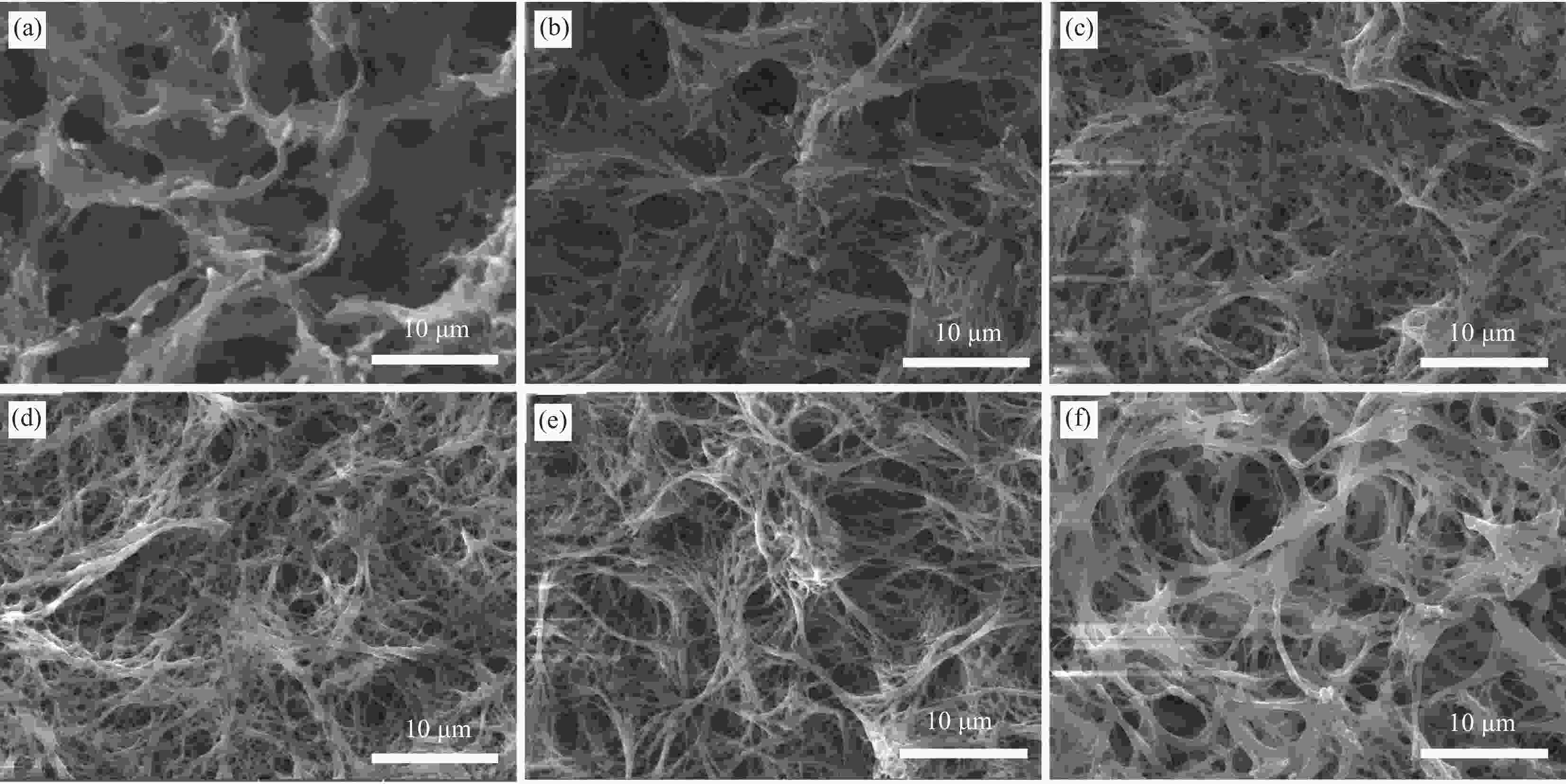
图 3 不同淬火时间PCL-CA-PLLA微-纳米复合纤维的SEM图像
Figure 3. SEM images of PCL-CA-PLLA composite micro-nanofibrous with different quenching time ((a) 1 min; (b) 3 min; (c) 5 min; (d) 10 min; (e) 30 min; (f) 60 min)
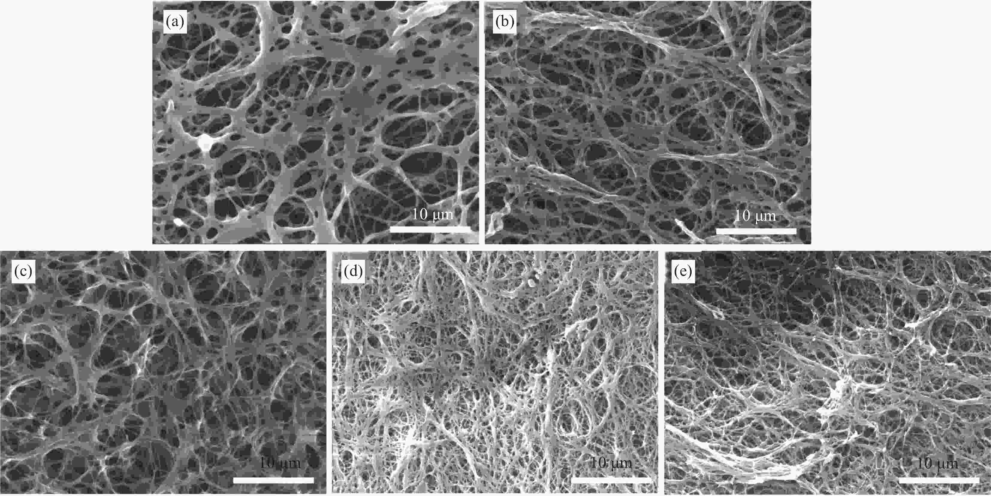
图 4 不同聚合物浓度PCL-CA-PLLA微-纳米复合纤维的SEM图像
Figure 4. SEM images of PCL-CA-PLLA composite micro-nanofibrous with different polymer concentrations ((a) 3%; (b) 5%; (c) 7%; (d) 9%; (e) 12%)
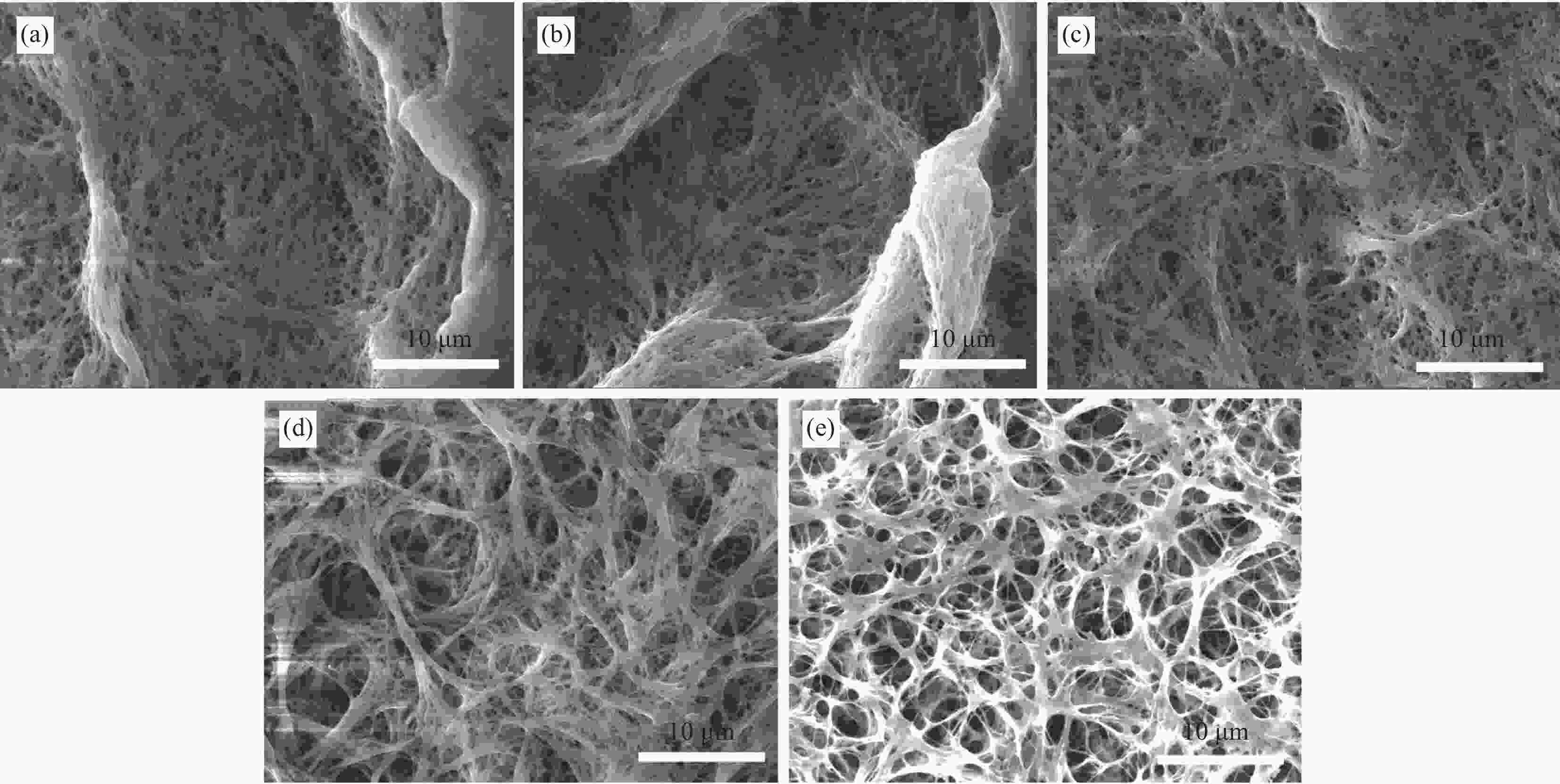
图 5 不同淬火温度PCL-CA-PLLA微-纳米复合纤维的SEM图像
Figure 5. SEM images of PCL-CA-PLLA composite micro-nanofibrous at different quenching temperatures((a) 10℃; (b) 0℃; (c) −10℃; (d) −20℃; (e) -30℃)
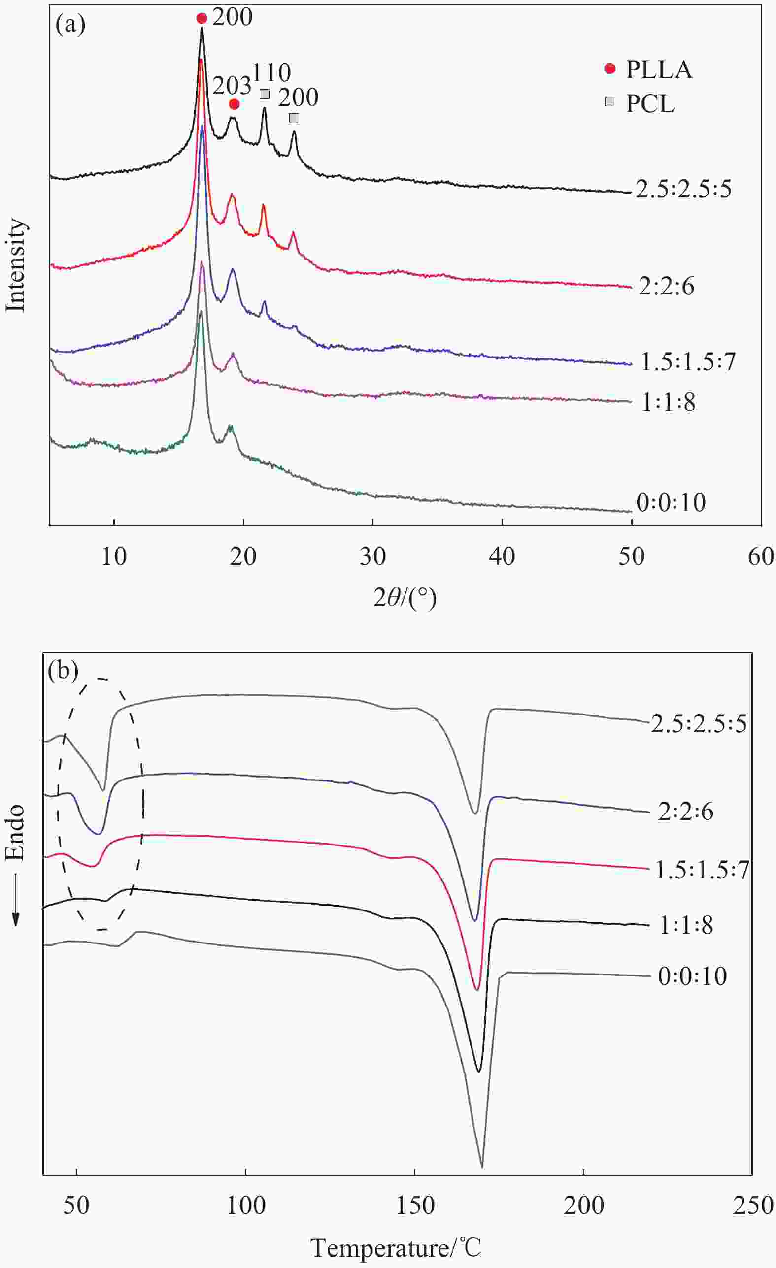
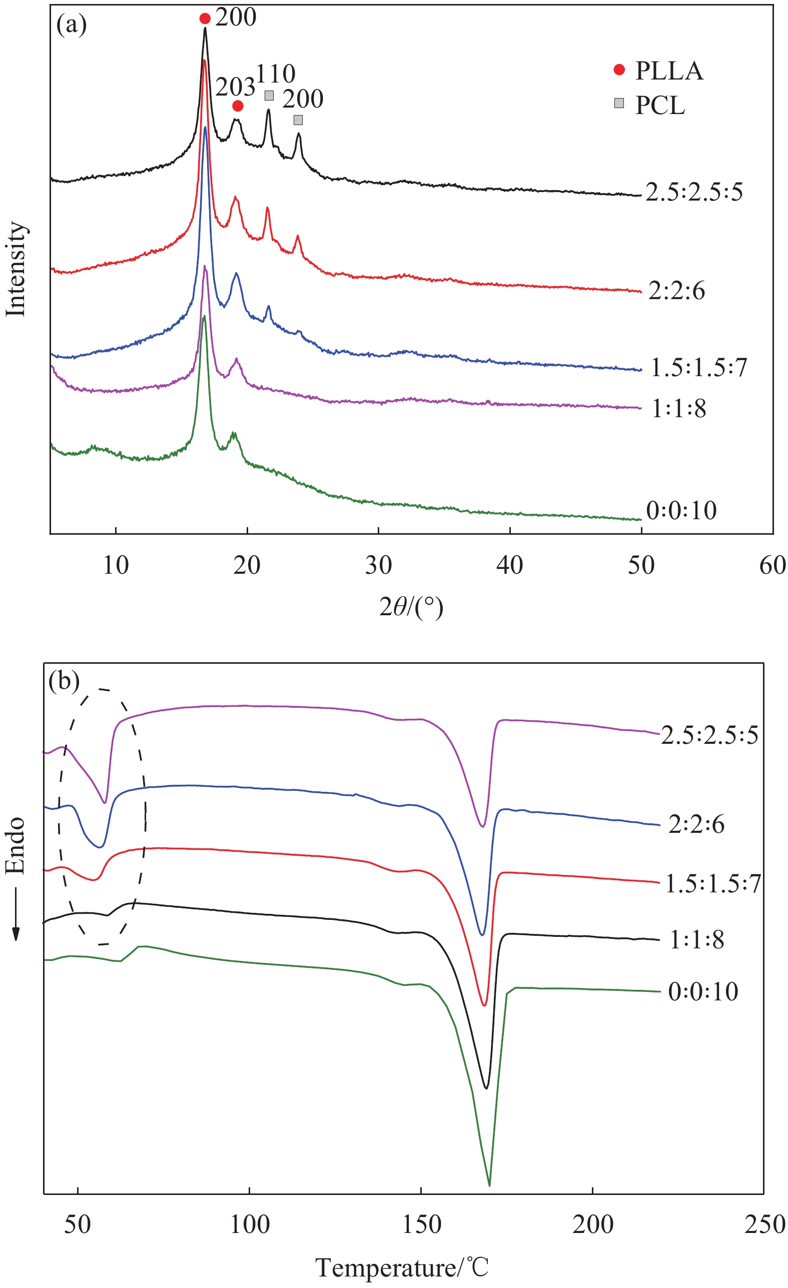
图 6 不同质量比PCL-CA-PLLA微-纳米复合纤维的XRE图谱 (a) 和DSC曲线 (b)
Figure 6. XRD patterns (a) and DSC curves (b) of PCL-CA-PLLA composite micro-nanofibrous with different mass ratios of PCL-CA-PLLA

图 7 PCL-CA-PLLA微-纳米复合纤维的N2吸附-脱附等温线 (a) 和孔容-孔径分布 (b)
Figure 7. N2 absorption-desorption isotherms (a) and pore size distribution curve (b) of PCL-CA-PLLA composite micro-nanofibrous

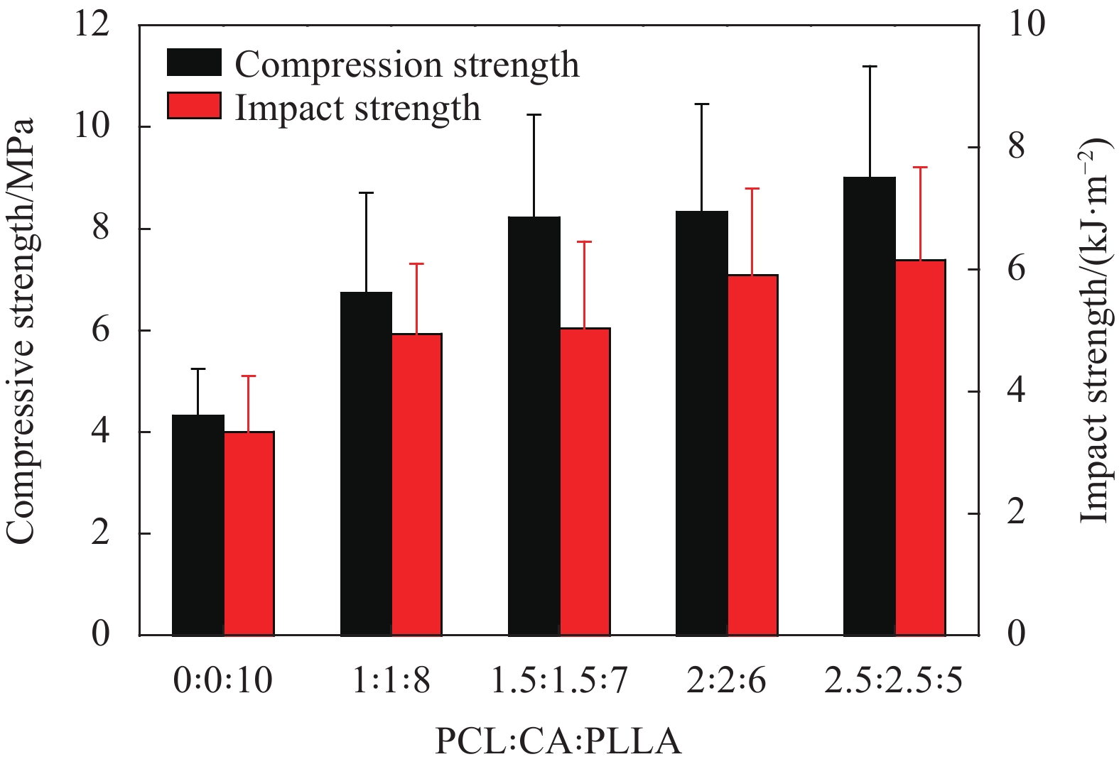
图 8 PCL-CA-PLLA微-纳米复合纤维压缩强度和冲击强度
Figure 8. Compressive strength and impact strength of PCL-CA-PLLA composite micro-nanofibrous
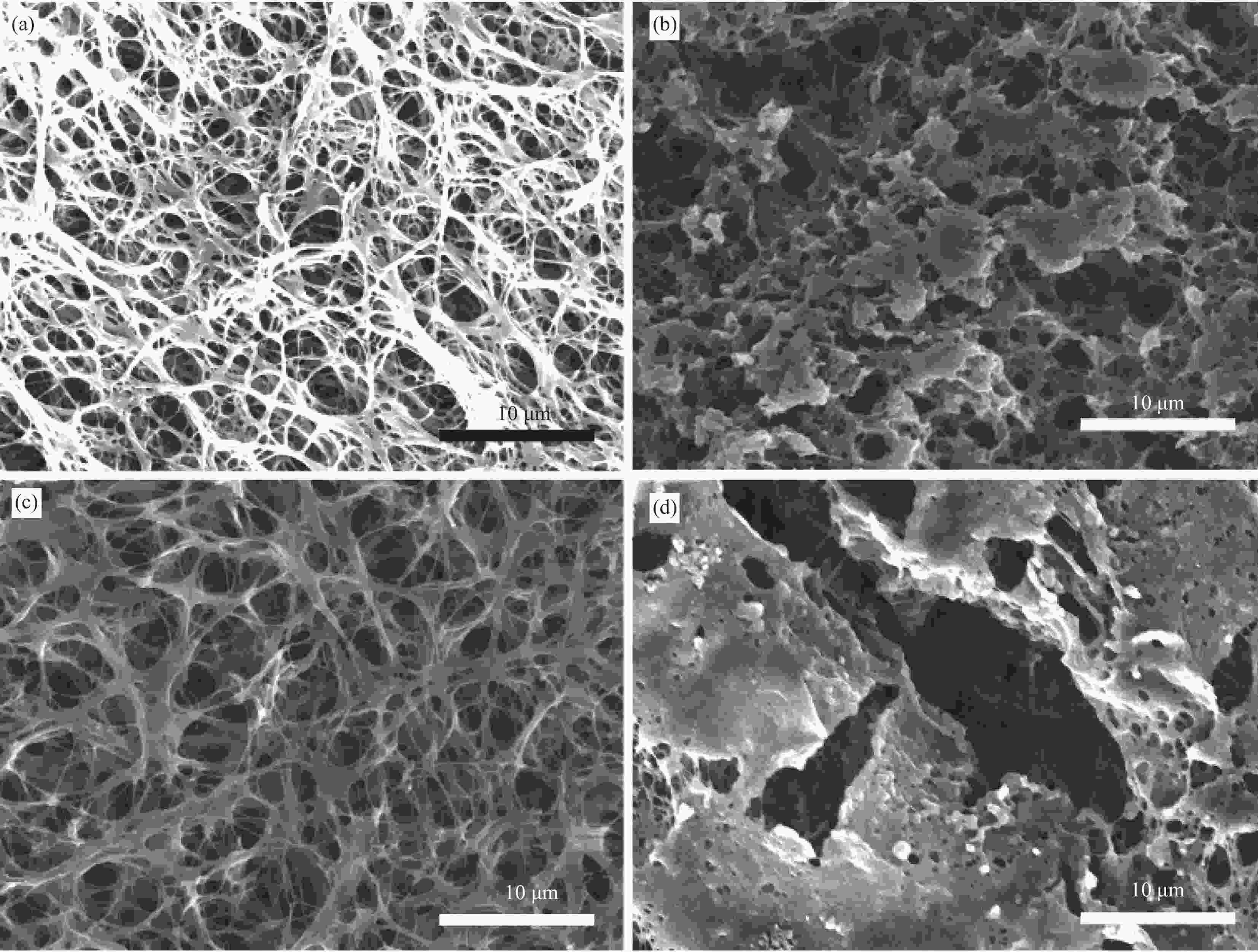
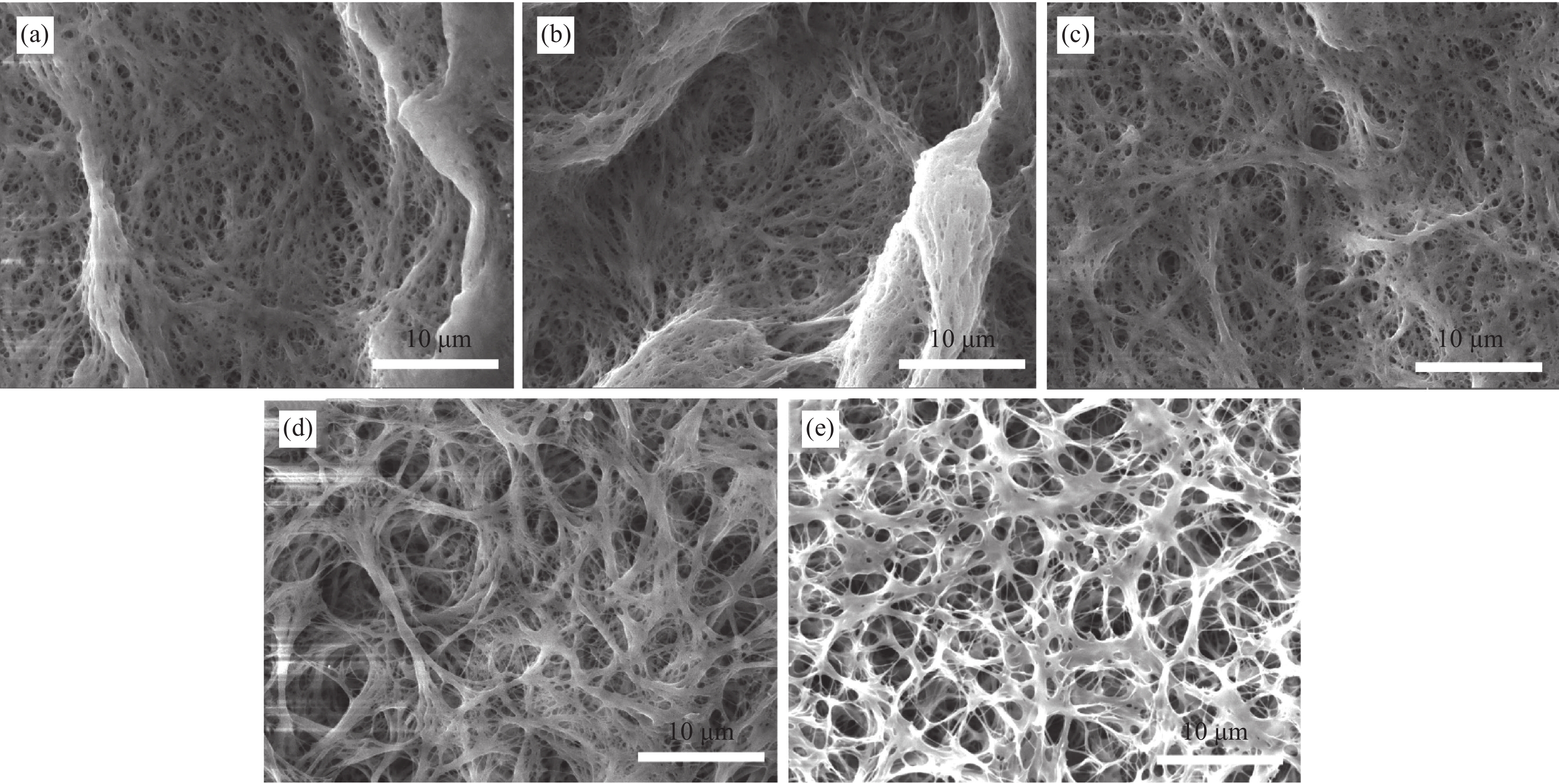
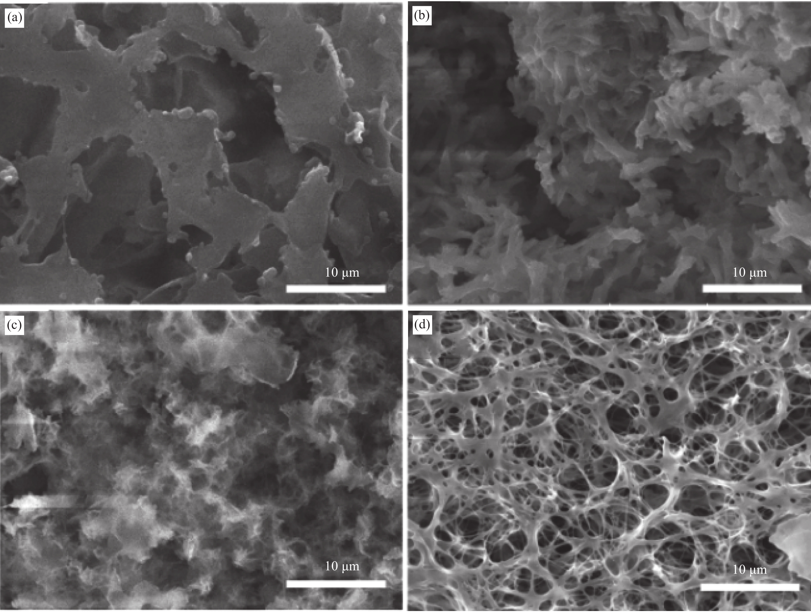
图 9 PCL-CA-PLLA微-纳米复合纤维支架浸泡在模拟体液(SBF)中14天的SEM图像
Figure 9. SEM images of PCL-CA-PLLA composite micro-nanofibrous scaffolds immersion in simulated body fluid (SBF) with 14 days ((a) Before immersion (PLLA); (b) After immersion (PLLA); (c) Before immersion (PCL-CA-PLLA); (d) After immersion (PCL-CA-PLLA))
表 1 左旋聚乳酸-聚己内酯-醋酸纤维素 (PCL-CA-PLLA)微-纳米纤维的制备条件
Table 1. Preparation condition of polycaprolactone-cellulose acetate-poly(L-lactic acid) (PCL-CA-PLLA) micro-nanofibrous
Experimental condition Solvent Mass ratio of PCL∶CA∶PLLA Concentration/wt% Quenching time/min Quenching temperature/℃ 1 DO, Acetonitrile,
DMAc, THF1∶1∶8 7 120 −30 2 THF 1∶1∶8, 1.5∶1.5∶7,
2∶2∶6, 2.5∶2.5∶57 120 −30 3 THF 1:1:8 7 1, 3, 5, 10, 30, 60 −30 4 THF 1:1:8 3, 5, 7, 9, 12 60 −30 5 THF 1∶1∶8 7 60 10, 0, −10, −20, −30 Notes: DO—1,4-dioxane; DMAc—N,N-dimethylacetamide; THF—Tetrahydrofuran.  下载: 导出CSV
表 2 溶剂和聚合物溶解度参数
Table 2. Solubility parameters of solvents and polymers
Solvent or polymer Solubility parameter/(J·cm−3)1/2 DO 20.3 Acetonitrile 24.5 DMAc 22.8 THF 18.5 PLLA 20.1 PCL 22.2 CA 21.1
下载: 导出CSV
表 3 PCL-CA-PLLA微-纳米复合纤维的热力学参数
Table 3. Thermal parameters of PCL-CA-PLLA composite micro-nanofibrous
Mass ratio of PCL∶CA∶PLLA Tm of PCL/℃ Tm of PLLA/℃ ΔHm of PCL/(J·g−1) Χc of PCL/% ΔHm of PLLA/(J·g−1) Χc of PLLA/% 0∶0∶10 − 169.80 − − −42.74 45.66 1∶1∶8 57.98 169.01 −4.88 35.10 −34.62 46.23 1.5∶1.5∶7 56.46 169.01 −7.23 34.67 −28.88 44.08 2∶2∶6 56.22 168.90 −10.13 36.43 −25.38 45.19 2.5∶2.5∶5 57.48 168.84 −12.66 36.43 −21.10 45.09 Notes: Tm—Melting temperature; ΔHm—Melting enthalpy; Χc—Degree of crystallinity.
下载: 导出CSV
表 4 PCL-CA-PLLA微-纳米复合纤维各物理参数
Table 4. Physics parameters of PCL-CA-PLLA composite micro-nanofibrous
Mass ratio of PCL∶CA∶PLLA Water contact angle/(°) Porosity/% Water absorption/% Specific surface area/(m2·g−1) 0∶0∶10 127.1±3.1 94.13 6.13 52.46 1∶1∶8 93.1±4.1 95.12 11.15 54.18 1.5∶1.5∶7 83.1±2.4 90.10 13.29 46.14 2∶2∶6 76.3±2.1 88.23 15.33 45.22 2.5∶2.5∶5 70.2±1.6 90.11 16.19 40.13
下载: 导出CSV
-
[1] FENG J, HUANG Z, DONG Y. Preparation of ice microspheres and their application in the preparation of porous poly (L-lactic acid)(PLLA) scaffolds[J]. Journal of Mater-ials Science,2019,54(4):3661-3670. doi: 10.1007/s10853-018-3086-6 [2] 廖欣宇, 王福科, 王国梁. 骨组织工程支架的进展与挑战[J]. 中国组织工程研究, 2021, 25(28):4553-4560.LIAO Xinyu, WANG Fuke, WANG Guoliang. Progress and challenges of bone tissue engineering scaffolds[J]. Chinese Journal of Tissue Engineering Research,2021,25(28):4553-4560(in Chinese). [3] AKBARZADEH R, YOUSEFI A M. Effects of processing parameters in thermally induced phase separation technique on porous architecture of scaffolds for bone tissue engineering[J]. Journal of Biomedical Materials Research Part B: Applied Biomaterials,2014,102(6):1304-1315. doi: 10.1002/jbm.b.33101 [4] SMITH L A, LIU X, MA P X. Tissue engineering with nano-fibrous scaffolds[J]. Soft Matter,2008,4(11):2144-2149. doi: 10.1039/b807088c [5] WU S, LIU X, YEUNG K W K, et al. Biomimetic porous scaffolds for bone tissue engineering[J]. Materials Science & Engineering R Reports,2014,80:1-36. [6] TANG D, TARE R S, YANG L Y, et al. Biofabrication of bone tissue: approaches, challenges and translation for bone regeneration[J]. Biomaterials,2016,83:363-382. doi: 10.1016/j.biomaterials.2016.01.024 [7] RUSHA A, MADHUMITHA A, PEARL A, et al. Biomaterials and cells for cardiac tissue engineering: Current choices[J]. Materials Science & Engineering C,2017,79:950-957. [8] ROSETI L, PARISI V, PETRETTA M, et al. Scaffolds for bone tissue engineering: State of the art and new perspectives[J]. Materials Science & Engineering,2017,78(9):1246-1262. [9] SILL T J. Electrospinnning : Applications in drug delivery and tissue engineering[J]. Biomaterials,2008,29(13):1989-2006. doi: 10.1016/j.biomaterials.2008.01.011 [10] LI J, CHEN Y, MAK A, et al. A one-step method to fabricate PLLA scaffolds with deposition of bioactive hydroxyapatite and collagen using ice-based microporogens[J]. Acta Biomaterialia,2010,6(6):2013-2019. doi: 10.1016/j.actbio.2009.12.008 [11] LIN L, HU Q, HUANG X, et al. Design and fabrication of bone tissue engineering scaffolds via rapid prototyping and CAD[J]. Journal of Rare Earths,2007,25(2):379-383. [12] RAEISDASTEH H V, DAVARAN S, RAMAZANI A, et al. Design and fabrication of porous biodegradable scaffolds: A strategy for tissue engineering[J]. Journal of Biomaterials Science: Polymer Edtion,2017,28(16):1-47. [13] 刘淑琼, 吴芳芳, 刘瑞来. 聚乳酸/聚乙烯吡咯烷酮纳米纤维复合支架的制备及性能[J]. 高分子材料科学与工程, 2015(6):172-176.LIU Shuqiong, WU Fangfang, LIU Ruilai. Preparation and property of polylactide/polyvinylpyrrolidone composite nanofibrous scaffolds[J]. Polymer Materials Science and Engineering,2015(6):172-176(in Chinese). [14] CONOSCENTI G, SCHNEIDER T, STOELZEL K, et al. PLLA scaffolds produced by thermally induced phase separation (TIPS) allow human chondrocyte growth and extracellular matrix formation dependent on pore size[J]. Materials Science & Engineering C,2017,80(3):449-459. [15] HOKMADAD V R, DAVARAN S, RAMZANI A, et al. Design and fabrication of porous biodegradable scaffolds: A strategy for tissue engineering[J]. Journal of Biomaterials Science: Polymer Edition,2017,28(16):1797-1825. doi: 10.1080/09205063.2017.1354674 [16] ONDER O C, YILGOR E, YILGOR I. Preparation of monolithic polycaprolactone foams with controlled morph-ology[J]. Polymer,2018,136(4):166-178. [17] UZ M, BUYUKOZ M, SHARMA A D, et al. Gelatin-based 3D conduits for transdifferentiation of mesenchymal stem cells into Schwann cell-like phenotypes[J]. Acta Biomater-ialia,2017,53(4):293-306. [18] WANG W, MIAO Y, ZHOU X, et al. Local delivery of BMP-2 from poly(lactic-co-glycolic acid) microspheres incorporated into porous nanofibrous scaffold for bone tissue regeneration[J]. Journal of Biomedical Nanotechnology,2017,13(11):1446-1456. doi: 10.1166/jbn.2017.2445 [19] KIM G M, LE K H T, GIANITELLI S M, et al. Electrospinning of PCL/PVP blends for tissue engineering scaffolds[J]. Journal of Materials Science: Materials in Medicine,2013,24(6):1425-1442. doi: 10.1007/s10856-013-4893-6 [20] KOUYA T, TADA S I, MINBU H, et al. Microporous membranes of PLLA/PCL blends for periosteal tissue scaffold[J]. Materials Letters,2013,95(3):103-106. [21] LIU S, ZHENG Y, HU J, et al. Fabrication and characterization of polylactic acid/polycaprolactone composite macroporous micro-nanofiber scaffolds by phase separation[J]. New Journal of Chemistry,2020,44(40):17382-17390. doi: 10.1039/D0NJ03176C [22] HOU J Z, SUN X P, ZHANG W X, et al. Preparation and characterization of electrospun fibers based on poly(L-lactic acid)/cellulose acetate[J]. Chinese Journal of Polymer Science,2012,30(6):916-922. doi: 10.1007/s10118-012-1191-6 [23] LIU R L, LIU J S, LIU H Q. Fabrication of PLLA/CA compo-sites porous ultrafine fibers[J]. Acta Polymerica Sinica,2013(10):1312-1318. [24] LIU R L, LI K N, LIU M, et al. Free poly(L-lactic acid) spherulites grown from thermally induced phase separation and crystallization kinetics[J]. Journal of Polymer Science Part B: Polymer Physics,2014,52(22):1476-1489. doi: 10.1002/polb.23587 [25] HE L M, ZHANG Y Q. Fabrication and characterization of poly (L-lactic acid) 3D nanofibrous scaffolds with controlled architecture by liquid-liquid phase separation from a ternary polymer-solvent system[J]. Polymer,2009,50(16):4128-4138. doi: 10.1016/j.polymer.2009.06.025 [26] GUARINO V, TADDEI P, FOGGIA M D, et al. The influence of hydroxyapatite particles on in vitro degradation behavior of poly ɛ-caprolactone-based composite scaffolds[J]. Tissue Engineering Part A,2009,15(11):3655-3668. doi: 10.1089/ten.tea.2008.0543 [27] CUNETY T A. Synthesis of biomimetic Ca-hydroxyapatite powders at 37℃ in synthetic body fluids[J]. Biomaterials,2000,21(14):1429-1438. doi: 10.1016/S0142-9612(00)00019-3 [28] 刘瑞来. 聚乳酸纳米纤维球晶多孔材料的制备及其结构性能研究[D]. 福州: 福建师范大学, 2015.LIU Ruilai. Fabrication, properties and application of porous materials based on PLLA spherulites composed of nano-fibers[D]. Fuzhou: Fujian Normal University, 2015(in Chinese). [29] SING K S W. Reporting physisorption data for gas/solid systems with special reference to the determination of surface area and porosity[J]. Pure and Applied Chemistry,1985,57(4):603-619. doi: 10.1351/pac198557040603 [30] 刘淑琼, 肖秀峰. PVP/PCL纳米纤维复合支架的生物活性[J]. 材料研究学报, 2014, 28(10):769-774. doi: 10.11901/1005.3093.2014.213LIU Shuqiong, XIAO Xiufeng. Cytocompatibility of PCL/PVP composite nanofibrous scaffolds[J]. Chinese Journal of Materials Research,2014,28(10):769-774(in Chinese). doi: 10.11901/1005.3093.2014.213 [31] LIU S, ZHENG Y, LIU R, et al. Preparation and characterization of a novel polylactic acid/hydroxyapatite composite scaffold with biomimetic micro-nanofibrous porous structure[J]. Journal of Materials Science: Materials in Medicine,2020,31(8):1-11. -

 下载:
下载:


 点击查看大图
计量
- 文章访问数: 974
- HTML全文浏览量: 372
- PDF下载量: 23
- 被引次数: 0
