


Bacteriostatic study of Kanamycin synergistic Cu2O/CuO composites targeting the bacterial cell wall
-
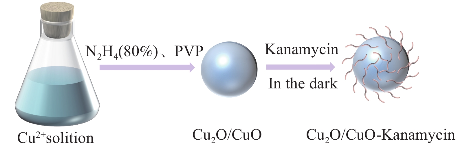
摘要: 随着耐药菌的出现,以传统抗生素为代表的抑菌剂药用价值逐步降低,因此,急需开发新型抗菌剂来解决细菌耐药和提升抗生素药效问题。本文用[Cu(NO3)2·3H2O]和N2H4·H2O制备纳米氧化亚铜(Cu2O/CuO),最后通过“浸渍法”将硫酸卡那霉素(Kanamycin)负载到纳米氧化亚铜上得到卡那霉素协同Cu2O/CuO (Cu2O/CuO-Kanamycin)纳米复合材料,并对Cu2O/CuO-Kanamycin复合材料的形貌结构、元素含量和键合方式等进行系统表征。以模型菌革兰氏阴性菌大肠杆菌(E. coli)、革兰氏阳性菌金黄色葡萄球菌 (S. aureus)和耐卡那霉素-沙门氏菌(D-Salm)为对象研究Cu2O/CuO-Kanamycin复合材料的抑菌效率及其作用机制。表征结果显示,制备的实心立方体Cu2O/CuO结构,因与空气有较小的接触面积而相对稳定,可与Kanamycin的—OH基团相互吸引并发生配位键合。抑菌活性表明,Cu2O/CuO-Kanamycin复合物在50 µg/mL浓度下,20 min内对E. coli、S. aureus和D-Salm的抑菌率超过99%,其中对E. coli敏感性更高。抑菌机制证明,复合材料主要通过破坏细菌细胞壁的结构而使细菌死亡。此研究不仅可提升传统抗生素的药用价值,且对耐药菌的抗菌性能显著提高,同时为医疗材料和环境卫生等领域提供广泛的科学依据。Abstract: With the emergence of drug-resistant bacteria, the medical value of bacteriostatic agents represented by traditional antibiotics is gradually decreasing, so there is an urgent need to develo novel antimicrobials to solve the problems of bacterial drug resistance and improve the efficacy of antibiotics. In this paper, we prepared cuprous oxide nanoparticles (Cu2O/CuO) from [Cu(NO3)2·3H2O] and N2H4·H2O, and then loaded kanamycin sulfate onto the cuprous oxide nanoparticles by the impregnation method. Finally, kanamycin sulfate (Kanamycin) was loaded onto the cuprous oxide nanocomposites by the "impregnation method" to obtain Kanamycin-coordinated Cu2O/CuO (Cu2O/CuO-Kanamycin) nanocomposites, and theMorphological structure, elemental content and bonding mode of the Cu2O/CuO-Kanamycin composites were systematically characterised. Gram-negative Escherichia coli (E. coli), Gram-positive Staphylococcus aureus (S. aureus) and Kanamycin-resistant Salmonella (D-Salm) were used as model organisms to study the bacterial inhibition efficiency of Cu2O/CuO-Kanamycin composites and their mechanism of action. The characterisation results showed that the prepared solid cubic Cu2O/CuO structure, which is relatively stable due to the small contact area with air, can be mutually attracted and ligated with the —OH group of kanamycin. The inhibitory activities showed that the Cu2O/CuO kanamycin complexes exhibited more than 99% inhibition of E. coli, S. aureus and D-Salm within 20 min at a concentration of 50 µg/ml, with a higher susceptibility to E. coli. The mechanism of bacterial inhibition showed that the composites killed the bacteria mainly by disrupting the structure of the bacterial cell wall. This study not only enhances the medicinal value of traditional antibiotics and significantly improves the antimicrobial performance against drug-resistant bacteria, but also provides a broad scientific basis for the fields of medical materials and environmental hygiene.
-
Key words:
- Cuprous oxide /
- Kanamycin sulfate /
- Synergistic bacteriostasis
-

图 3 Cu2O/CuO和Cu2O/CuO-Kanamycin复合材料的X射线衍射图谱(a);Kanamycin、Cu2O/CuO和Cu2O/CuO-Kanamycin复合材料的傅立叶变换红外光谱图(b)
Figure 3. The XRD of Cu2O/CuO and Cu2O/CuO-Kanamycin composites (a); Fourier transform infrared spectra of Kanamycin, Cu2O/CuO and Cu2O/CuO-Kanamycin composites (b)

图 4 纳米复合材料的XPS总谱(a)。Cu 2 p (b)、C 1 s (c)、O 1 s (d)、N 1 s (e)、S 2 p (f)的XPS光谱
Figure 4. XPS gross spectra of nanocomposites (a). XPS spectra of Cu 2 p (b), C 1 s (c), O 1 s (d), N 1 s (e), S 2 p (f)

图 5 Cu2O与硫酸卡那霉素(Kanamycin)的电子结构优化及ESP分析图(a);Cu2O与Kanamycin的结合能分析图(b)
Figure 5. The objective of this study was to optimise the electronic structure of Cu2O in the presence of Kanamycin sulphate (Kanamycin) and to analyse the binding energy of Cu2O with Kanamycin

图 6 Kanamycin、Cu2O/CuO和Cu2O/CuO-Kanamycin复合材料对大肠杆菌 (a)、金黄色葡萄球菌(b)和耐卡那霉素-沙门氏菌(c)的抑菌圈直径曲线
Figure 6. The inhibition diameter profiles of Kanamycin, Cu2O/CuO and Cu2O/CuO-Kanamycin composites against Escherichia coli (a), Staphylococcus aureus (b) and Kanamycin-resistant Salmonella (c) are presented in the following figures

图 7 Cu2O/CuO-Kanamycin复合材料对大肠杆菌 (a)、金黄色葡萄球菌(b)和耐卡那霉素-沙门氏菌(c)的菌落计数结果;Cu2O/CuO-Kanamycin复合材料在不同时间对大肠杆菌、金黄色葡萄球菌和耐卡那霉素-沙门氏菌的抑菌率(d)和菌落数(e)
Figure 7. The results of colony counting for Cu2O/CuO-Kanamycin composites against Escherichia coli (a), Staphylococcus aureus (b) and Kanamycin-resistant Salmonella (c) are presented. Inhibition rate (d) and number of colonies (e) of Cu2O/CuO-Kanamycin composites against Escherichia coli, Staphylococcus aureus and Kanamycin-resistant Salmonella at various times are also shown

图 8 Zeta电势分析结果(a)。Cu2O/CuO-Kanamycin复合材料与大肠杆菌(b)、金黄色葡萄球菌(c)和耐卡那霉素-沙门氏菌(d)作用后细胞质泄露实验结果
Figure 8. Results of Zeta potential analysis (a). Results of cytoplasmic leakage experiments after the action of Cu2O/CuO-Kanamycin composites with Escherichia coli (b), Staphylococcus aureus (c) and Kanamycin-resistant Salmonella (d)

图 9 Cu2O/CuO-Kanamycin纳米复合材料对大肠杆菌(d)、金黄色葡萄球菌(e)和耐药卡那霉素-沙门氏菌(f)的PI染色结果。(a、b、c)为对应的纯菌效果图
Figure 9. PI staining results of Cu2O/CuO-Kanamycin nanocomposites against E. coli (d), S. aureus (e) and D-Salm (f). (a, b, c) are the corresponding pure bacterial results

图 10 Kanamycin协同Cu2O/CuO的抑菌机制流程图
Figure 10. Flowchart of the inhibition mechanism of Kanamycin synergising with Cu2O/CuO
表 1 Cu2O/CuO-Kanamycin复合材料对大肠杆菌、金黄色葡萄球菌和耐卡那霉素-沙门氏菌的MIC值
Table 1. MIC values of Cu2O/CuO-Kanamycin composites against E. coli, S. aureus and D-Salm.
Beta-bacteria Material/ Material Concentration Gradientc ( µg/mL )/ Colony Concentration (1×10⁵ CFU/mL) Cu2O/CuO-Kanamycin composite material 0 10 20 30 40 50 60 70 E. coli 7.44 6.17 3.81 1.11 0.21 0.21 0.19 0.17 S. aureus 5.31 3.15 2.37 2.23 0.18 0.18 0.15 0.14 D-Salm 6.89 5.41 3.78 2.14 0.23 0.18 0.15 0.13  下载: 导出CSV
-
[1] HANADA S, PIRZADEH M, CARVER K Y, et al. Respiratory viral infection-induced microbiome alterations and secondary bacterial pneumonia[J]. Frontiers in immunology, 2018, 9: 2640. doi: 10.3389/fimmu.2018.02640 [2] 钟艾玲, 田敏, 刘艳全等. 氨基糖苷类抗生素的耐药机制研究进展[J]. 中国抗生素杂志, 2019, 44(4): 401-405. doi: 10.3969/j.issn.1001-8689.2019.04.002ZHONG A L, TIAN M, LIU Y et al. Progress of resistance mechanism of aminoglycoside antibiotics[J]. Chinese Journal of Antibiotics, 2019, 44(4): 401-405(in Chinese). doi: 10.3969/j.issn.1001-8689.2019.04.002 [3] 张小敏, 郭全友, 周国燕等. 壳聚糖协同乳酸链球菌素抗轻腌大黄鱼源特定腐败菌抑制效应研究[J]. 中国食品学报, 2022, 22(5): 259-270.ZHANG X M, GUO Q Y, ZHOU G Y et al. Chitosan synergises with streptozotocin lactate to inhibit specific spoilage organisms from lightly pickled rhubarb fish[J]. Chinese Journal of Food Science, 2022, 22(5): 259-270(in Chinese). [4] GAO J, Yan Y, GAO S, et al. Heterogeneous Cu2O-SnO2 doped polydopamine fenton-like nanoenzymes for synergetic photothermal-chemodynamic antibacterial application[J]. Acta Biomaterialia, 2024, 173: 420-431. doi: 10.1016/j.actbio.2023.11.009 [5] BOST M, HOUDART S, OBERLI M, et al. Dietary copper and human health: Current evidence and unresolved issues[J]. Journal of trace elements in medicine and biology, 2016, 35: 107-115. doi: 10.1016/j.jtemb.2016.02.006 [6] DALECKI A G, CRAWFORD C L, WOLSCHENDORF F. Copper and antibiotics: discovery, modes of action, and opportunities for medicinal applications[J]. Advances in microbial physiology, 2017, 70: 193-260. [7] PATWARDHAN A, COWAN J A. Influence of charge and structure on the coordination chemistry of copper aminoglycosides[J]. Dalton Transactions, 2011, 40(8): 1795-1801. doi: 10.1039/c0dt00704h [8] MANNING T, PATEL H, WYLIE G, et al. Structural measurements and cell line studies of the copper-PEG-Amikacin complex against Mycobacterium tuberculosis[J]. Bioorganic & Medicinal Chemistry Letters, 2015, 25(24): 5825-5830. [9] VARAPRASAD K, LÓPEZ M, NÚÑEZ D, et al. Antibiotic copper oxide-curcumin nanomaterials for antibacterial applications[J]. Journal of Molecular Liquids, 2020, 300: 112353. doi: 10.1016/j.molliq.2019.112353 [10] 吴迎花, 陈惠惠, 房迅等. Cu2O/CuO-四环素复合材料的协同抑菌性能[J]. Acta Materiae Compositae Sinica, 2023, 40(12).WU Y H, CHEN H H, FANG X et al. Synergistic antibacterial properties of Cu2O/CuO-tetracycline composites[J]. Acta Materiae Compositae Sinica, 2023, 40(12). (in Chinese) [11] PU J, ZHANG Z, ZHANG H, et al. Efficacy of Bactericides Against Potato Common Scab Caused by Streptomyces in Yunnan, China[J]. American Journal of Potato Research, 2022, 99(4): 326-335. doi: 10.1007/s12230-022-09883-2 [12] WEI C, FAN C, XIE D, et al. Fabrication of cinnamaldehyde-entrapped ethosome nanoparticles as antimicrobial agent[J]. LWT, 2023, 181: 114760. doi: 10.1016/j.lwt.2023.114760 [13] ÇETINKAYA E A, KOÇ A, KOÇ H K, et al. Synthesis, characterization and antimicrobial properties of silver complexes derived from 5, 6-Dimethylbenzimidazol-2-ylidene[J]. Polyhedron, 2023, 237: 116383. doi: 10.1016/j.poly.2023.116383 [14] MAILLARD A P V F, ESPECHE J C, Maturana P, et al. Zeta potential beyond materials science: Applications to bacterial systems and to the development of novel antimicrobials[J]. Biochimica et Biophysica Acta (BBA)-Biomembranes, 2021, 1863(6): 183597. doi: 10.1016/j.bbamem.2021.183597 [15] WANG J, FANG X, CHEN H, et al. Antibacterial properties of the flower shaped nano-CuFe2O4@ MoS2 composites[J]. Colloids and Surfaces A: Physicochemical and Engineering Aspects, 2024, 683: 133076. doi: 10.1016/j.colsurfa.2023.133076 [16] HE Q, TIAN Y, WU Y, et al. Electrochemical sensor for rapid and sensitive detection of tryptophan by a Cu2O nanoparticles-coated reduced graphene oxide nanocomposite[J]. Biomolecules, 2019, 9(5): 176. doi: 10.3390/biom9050176 [17] LIU S, ZHAO X, SUN H, et al. The degradation of tetracycline in a photo-electro-Fenton system[J]. Chemical Engineering Journal, 2013, 231: 441-448. doi: 10.1016/j.cej.2013.07.057 [18] TRIVEDI M K, PATIL S, SHETTIGAR H, et al. Spectroscopic characterization of chloramphenicol and tetracycline: An impact of biofield[J]. Pharm Anal Acta, 2015, 6(395): 19-21. [19] 檀苗苗, 丁娅莉, 山鹏禹等. Cu2O/PTCDI复合材料的制备及其光催化活性[J]. 化工环保, 2020, 40(3): 322-328. doi: 10.3969/j.issn.1006-1878.2020.03.016TAN M M, DING Y L, SHAN P Y et al. Preparation of Cu2O/PTCDI composites and their photocatalytic activity[J]. Chemical and Environmental Protection, Inc, 2020, 40(3): 322-328(in Chinese). doi: 10.3969/j.issn.1006-1878.2020.03.016 [20] WANG B, LI R, ZHANG Z, et al. Novel Au/Cu2O multi-shelled porous heterostructures for enhanced efficiency of photoelectrochemical water splitting[J]. Journal of Materials Chemistry A, 2017, 5(27): 14415-14421. doi: 10.1039/C7TA02254A [21] GHODSELAHI T, VESAGHI M A, SHAFIEKHANI A, et al. XPS study of the Cu@Cu2O core-shell nanoparticles[J]. Applied Surface Science, 2008, 255(5): 2730-2734. doi: 10.1016/j.apsusc.2008.08.110 [22] LI L, LEI J, JI T. Facile fabrication of p-n heterojunctions for Cu2O submicroparticles deposited on anatase TiO2 nanobelts[J]. Materials Research Bulletin, 2011, 46(11): 2084-2089. doi: 10.1016/j.materresbull.2011.06.033 [23] MAHMOUD M A, QIAN W, El-Sayed M A. Following charge separation on the nanoscale in Cu2O-Au nanoframe hollow nanoparticles[J]. Nano letters, 2011, 11(8): 3285-3289. doi: 10.1021/nl201642r [24] CHEN X, WANG X, FANG D. A review on C 1s XPS-spectra for some kinds of carbon materials[J]. Fullerenes, Nanotubes and Carbon Nanostructures, 2020, 28(12): 1048-1058. doi: 10.1080/1536383X.2020.1794851 [25] GUO H, CHENG J, MAO Y, et al. Fabricating different coordination states of cobalt as magnetic acid-base bifunctional catalyst for biodiesel production from microalgal lipid[J]. Fuel, 2022, 322: 124172. doi: 10.1016/j.fuel.2022.124172 [26] DOU H, XU M, ZHENG Y, et al. Bioinspired Tough Solid-State Electrolyte for Flexible Ultralong-Life Zinc-Air Battery[J]. Advanced Materials, 2022, 34(18): 2110585. doi: 10.1002/adma.202110585 [27] SIVKOV D V, PETROVA O V, NEKIPELOV S V, et al. Quantitative Characterization of Oxygen-Containing Groups on the Surface of Carbon Materials: XPS and NEXAFS Study[J]. Applied Sciences, 2022, 12(15): 7744. doi: 10.3390/app12157744 [28] WANG X L, LI Y, HUANG J, et al. Efficiency and mechanism of adsorption of low concentration uranium in water by extracellular polymeric substances[J]. Journal of environmental radioactivity, 2019, 197: 81-89. doi: 10.1016/j.jenvrad.2018.12.002 [29] ZHENG K, SETYAWATI M I, LIM T P, et al Antimicrobial cluster bombs: silver nanoclusters packed with daptomycin[J]. ACS nano, 2016, 10(8): 7934-7942. [30] FRANCO D, CALABRESE G, GUGLIELMINO S P P, et al. Metal-based nanoparticles: Antibacterial mechanisms and biomedical application[J]. Microorganisms, 2022, 10(9): 1778. doi: 10.3390/microorganisms10091778 [31] BERA S, ZHANEL G G, SCHWEIZER F. Antibacterial activities of aminoglycoside antibiotics-derived cationic amphiphiles. Polyol-modified neomycin B-, kanamycin A-, amikacin-, and neamine-based amphiphiles with potent broad spectrum antibacterial activity[J]. Journal of medicinal chemistry, 2010, 53(9): 3626-3631. doi: 10.1021/jm1000437 [32] GODOY-GALLARDO M, ECKHARD U, DELGADO L M, et al. Antibacterial approaches in tissue engineering using metal ions and nanoparticles: From mechanisms to applications[J]. Bioactive Materials, 2021, 6(12): 4470-4490. doi: 10.1016/j.bioactmat.2021.04.033 [33] CARRYN S, CHANTEUX H, SERAL C, et al. Intracellular pharmacodynamics of antibiotics[J]. Infectious Disease Clinics, 2003, 17(3): 615-634. [34] BREZDEN A, MOHAMED M F, NEPAL M, et al. Dual targeting of intracellular pathogenic bacteria with a cleavable conjugate of kanamycin and an antibacterial cell-penetrating peptide[J]. Journal of the American Chemical Society, 2016, 138(34): 10945-10949. doi: 10.1021/jacs.6b04831 -

 下载:
下载:


 点击查看大图
计量
- 文章访问数: 49
- HTML全文浏览量: 22
- 被引次数: 0
