


Preparation of multifunctional polyvinyl alcohol microspheres by electrospinning and its properties and application
-
摘要: 通过静电纺丝法,制备了MnO2-聚多巴胺(PDA)/聚乙烯醇(PVA)和MnO2-PDA-5-氟尿嘧啶(5-Fu)/PVA复合微球。具体地,在PVA溶液中依次加入多巴胺(DA)与KMnO4,在所得溶液中,DA与KMnO4发生氧化还原反应,原位聚合生成PDA,而KMnO4转变为MnO2,通过静电纺丝法制备了MnO2-PDA/PVA微球。向上述溶液中加入化疗药物5-Fu,通过静电纺丝法还可以制备MnO2-PDA-5-Fu/PVA微球。通过戊二醛交联赋予静电纺丝微球水稳定性。这一复合微球材料具有优异的光热转换性能、可控的药物缓释性能及良好的生物安全性,被应用于防止术后肿瘤的复发。一方面,在808 nm NIR激光照射下MnO2-PDA-5-Fu/PVA微球的光热转换效率可达到24.5%,实现高效的肿瘤热消融;另一方面,释放出来的5-Fu可杀死术后残余的肿瘤细胞。该研究论证了静电纺丝法制备复合微球的可行性,并证实了这种微球在防止肿瘤术后复发领域的应用潜力,为多功能复合微/纳米材料的设计提供了新策略。Abstract: In this paper, MnO2-polydopamine (PDA)/polyvinyl alcohol (PVA) and MnO2-PVA-5-fluorouracil (5-Fu)/PVA composite microspheres were prepared by electrospinning. Specifically, dopamine (DA) and KMnO4 were added to the PVA solution, where a redox reaction happened between DA and KMnO4 to form PDA and MnO2. Thus, the MnO2-PDA/PVA microspheres were prepared by electrospinning the MnO2-PDA/PVA solution. MnO2-PDA-5-Fu/PVA microspheres were prepared by electrospinning the MnO2-PDA-5-Fu/PVA solution, which was prepared by directly dissolving 5-Fu in the MnO2-PDA/PVA solution. The water stability of the electrospun microspheres was given by a glutaraldehyde crosslinking method. The composite microspheres have excellent photothermal conversion, controlled drug release and good biosafety, which were used to prevent the recurrence of tumor after the operation. The photothermal conversion efficiency of MnO2-PDA-5-Fu/PVA microspheres reaches 24.5% under the irradiation of 808 nm NIR laser, which can achieve the efficient tumor thermal ablation; on the other hand, the released 5-Fu can kill the residual tumor cells after operation. This study confirms the application potential of the microspheres in the field of preventing tumor recurrence, and provides a new strategy for the design of multifunctional composite nanomaterials.
-
Key words:
- electrospining /
- polyvinyl alcohol /
- polydopamine /
- photothermal therapy /
- tumor therapy
-

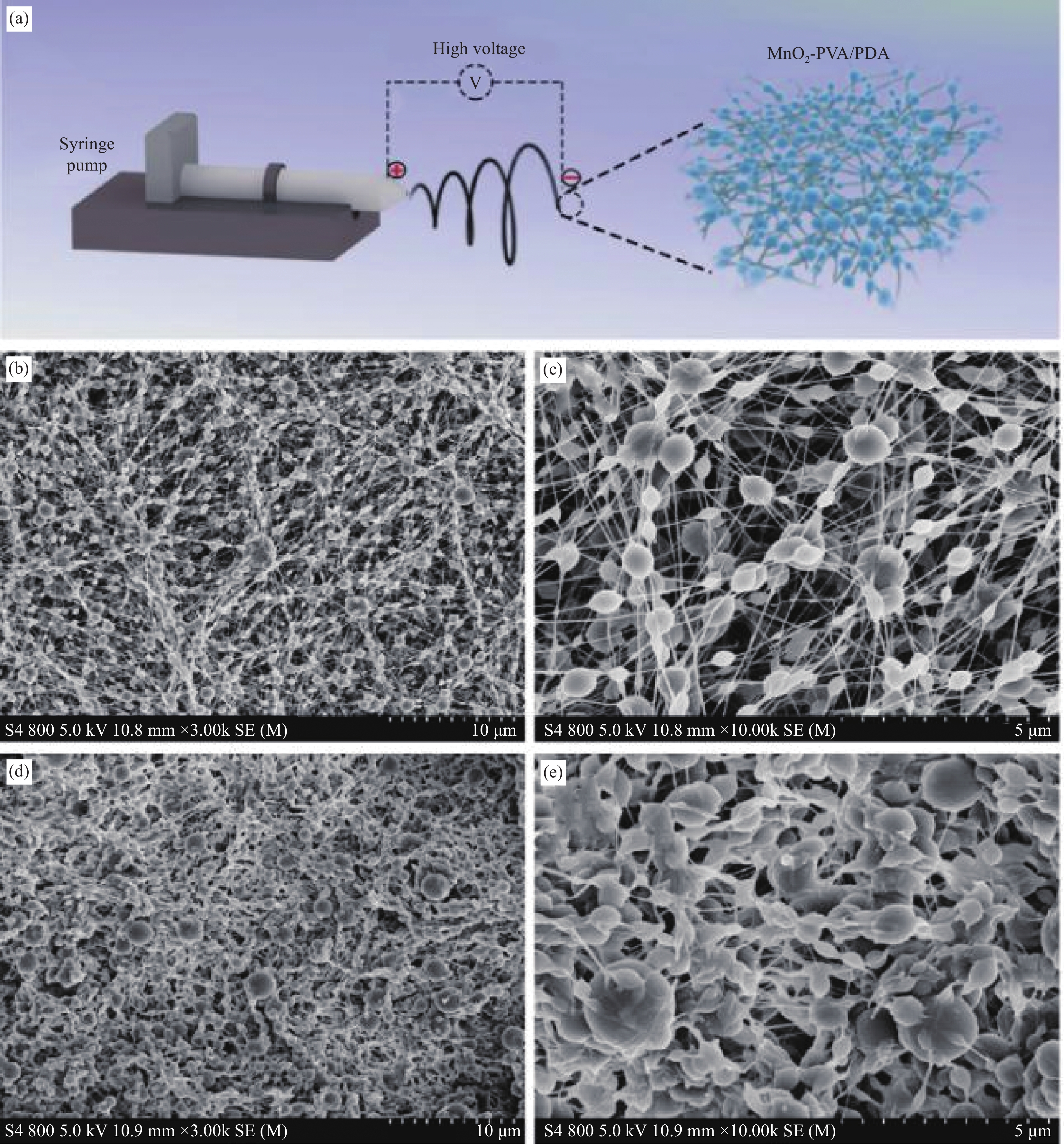
图 1 静电纺丝制备MnO2-聚多巴胺(PDA)/聚乙烯醇(PVA) 的示意图 (a)、未交联的MnO2-PDA/PVA (b) 和MnO2-PDA-5-氟尿嘧啶(5-Fu)/PVA (c) 的SEM图像、交联后的MnO2-PDA/PVA (d) 和MnO2-PDA-5-Fu/PVA (e) 的SEM图像
Figure 1. Schematic diagram of electrospinning MnO2-polydopamine (PDA)/polyvinyl alcohol (PVA) (a), SEM images of uncrosslinked MnO2-PD/PVA (b) and MnO2-PDA-5-fluorouracil (5-Fu)/PVA (c), SEM images of MnO2-PDA/PVA (d) and MnO2-PDA-5-Fu/PVA (e) after crosslinking
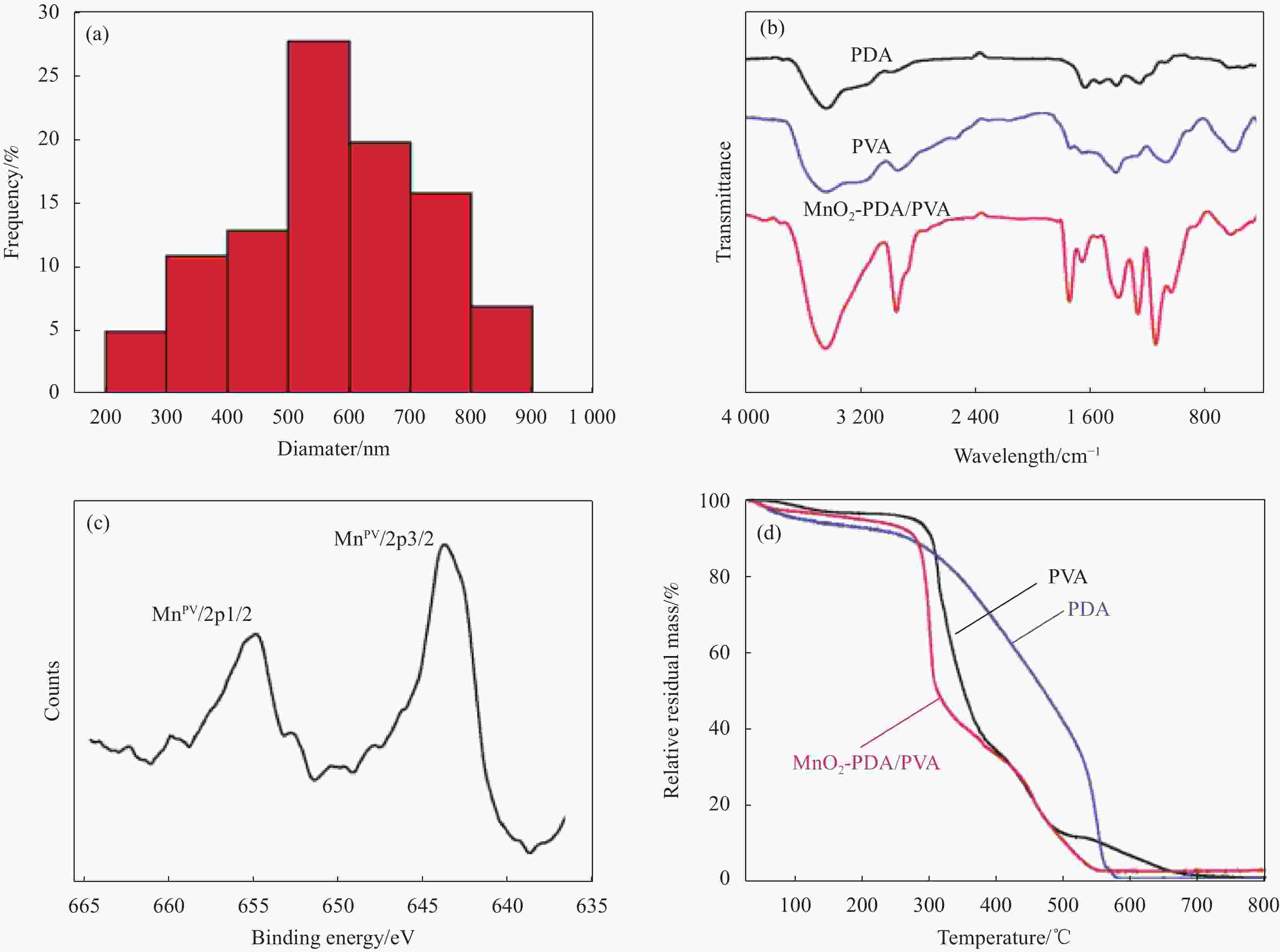
图 2 MnO2-PDA/PVA复合微球的粒径分布 (a)、MnO2-PDA/PVA的FTIR图谱 (b)、MnO2-PDA/PVA的XPS图谱 (c)、MnO2-PDA/PVA的热重曲线 (d)
Figure 2. Particle size distribution of the microspheres (a), FTIR spectra of different samples(b), XPS spectra of MnO2-PDA/PVA (c) , thermogravimetric curves of MnO2-PDA/PVA (d)

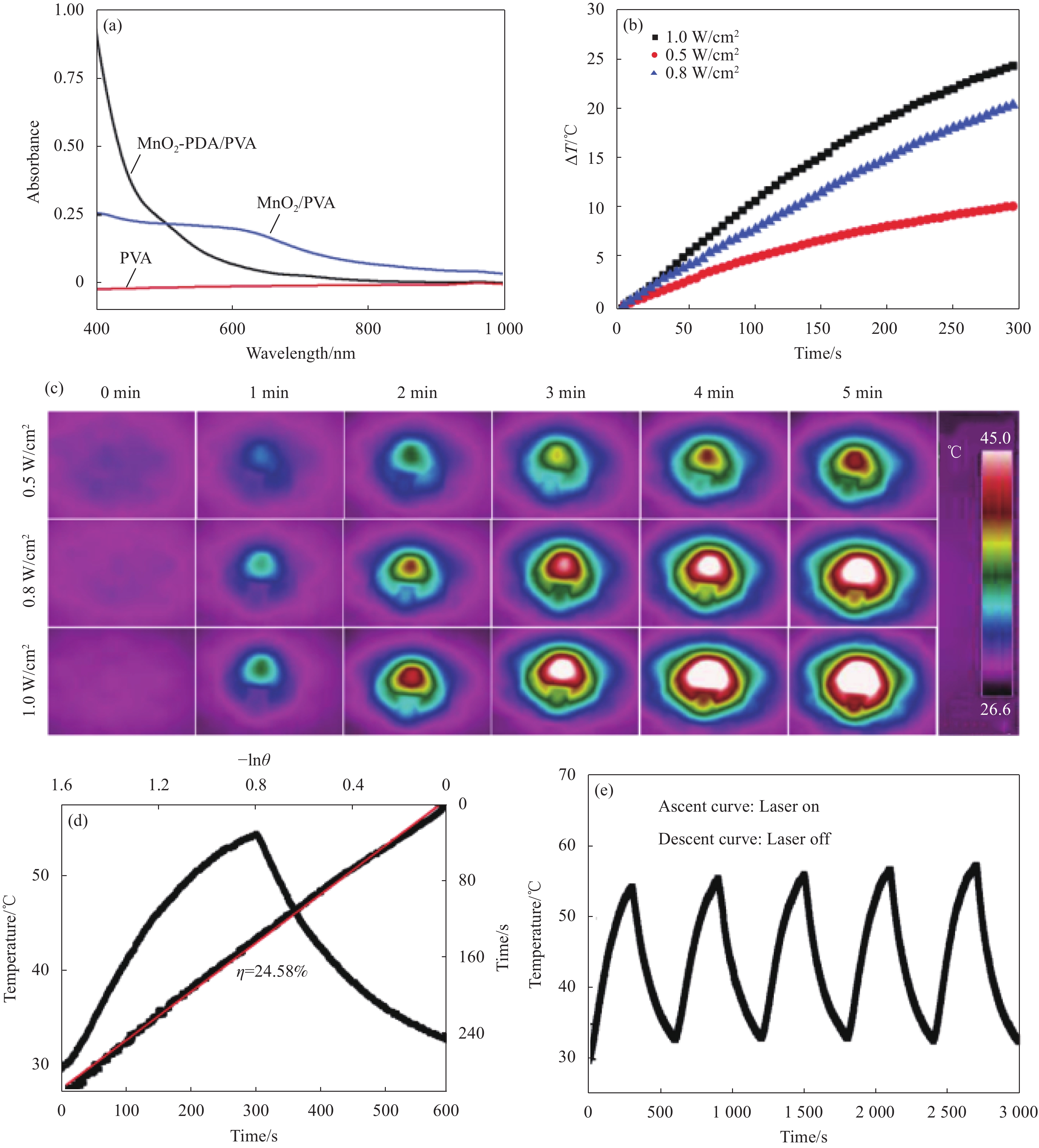
图 3 不同溶液的光吸收谱图 (a)、不同激光密度下微球的光热性能曲线 (b)、对应于图3(b)的光热成像照片 (c)、光热转换效率曲线 (d)、光热稳定性曲线 (e)
Figure 3. Light absorption of different solutions (a), photothermal performance curves under different laser densities (b), photothermal imaging photographs corresponding to fig. 3(b) (c), photothermal conversion efficiency curve (d), photothermal stability curve of MnO2-PVA/PDA (e)


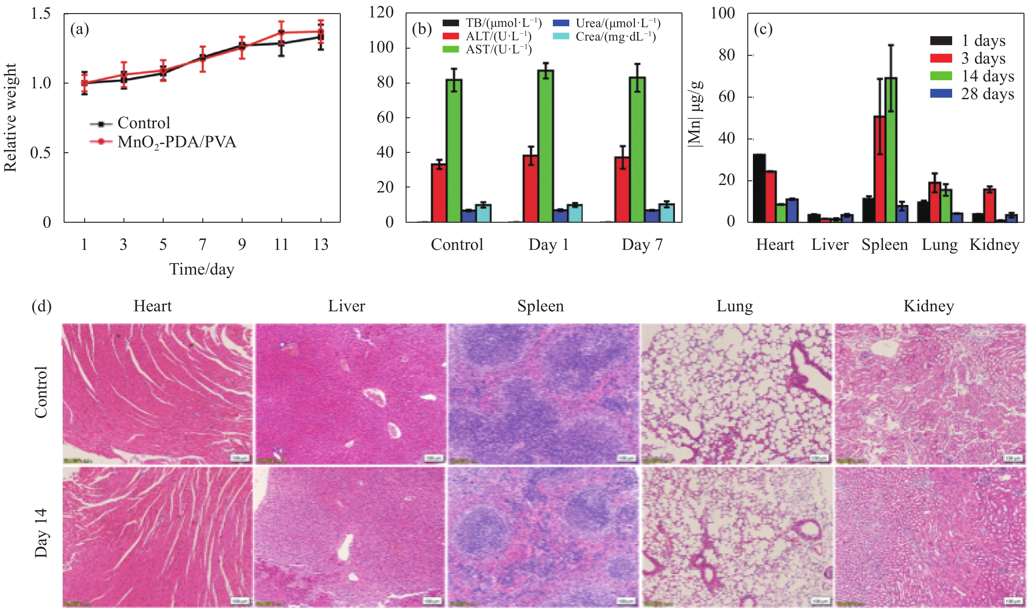
图 6 对照组小鼠和实验组小鼠在14天内的体重变化 (a)、血清生化因子 (b)、不同时间点小鼠主要器官中Mn元素的分布含量 (c)、小鼠主要器官的H&E染色 (d)
Figure 6. Body weight changes of control and experimental mice within 14 days (a), serum biochemical factors (b), distribution and content of Mn in main organs of mice at different time points (c), H&E staining of main organs of mice (d)
TB—Total bilirubin; ALT—Alanine transaminase; AST—Aspartate aminotransferase

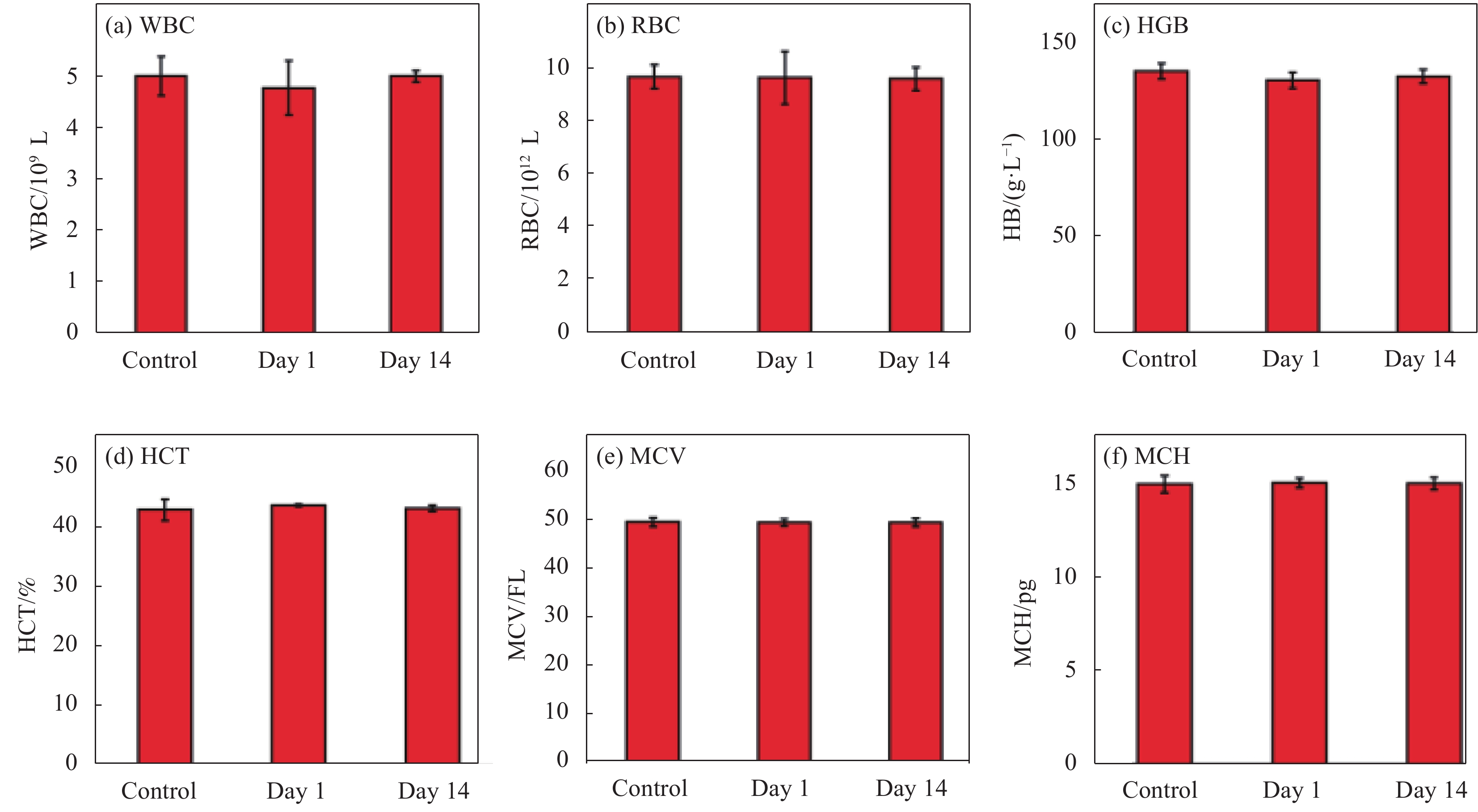
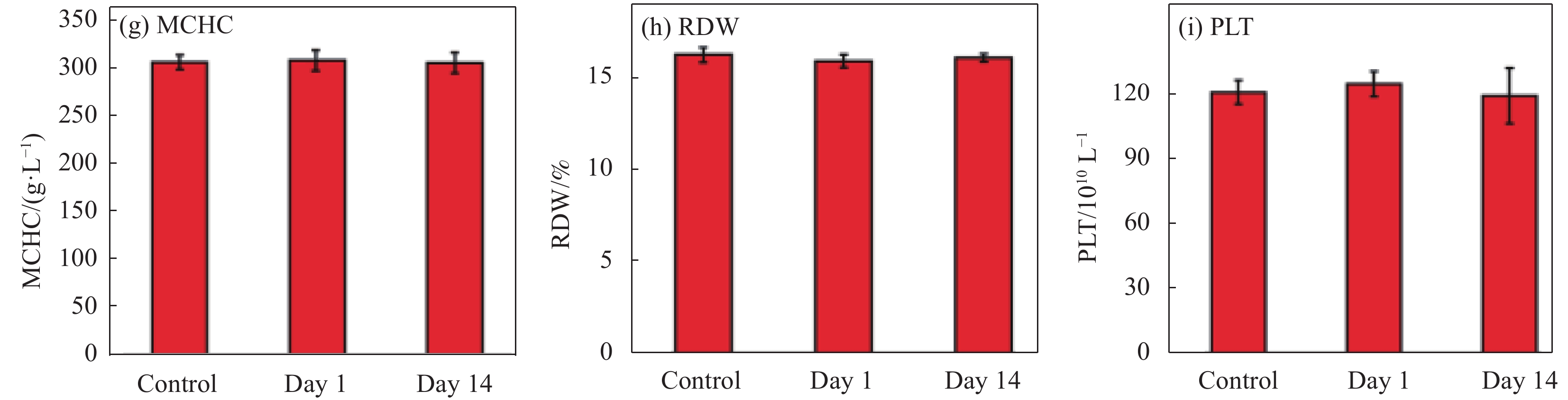
图 7 不同时间点的KM鼠血常规参数
Figure 7. Blood routine parameters of KM mice at different time points
WBC—White blood cell count; RBC—Red blood cell count; HB—Hemoglobin; HCT—Hematocrit; MCV—Corpuscular volume; MCH—Corpuscular hemoglobin; MCHC—Corpuscular hemoglobin concentration; RDW—Red cell distribution width; PLT—Platelet

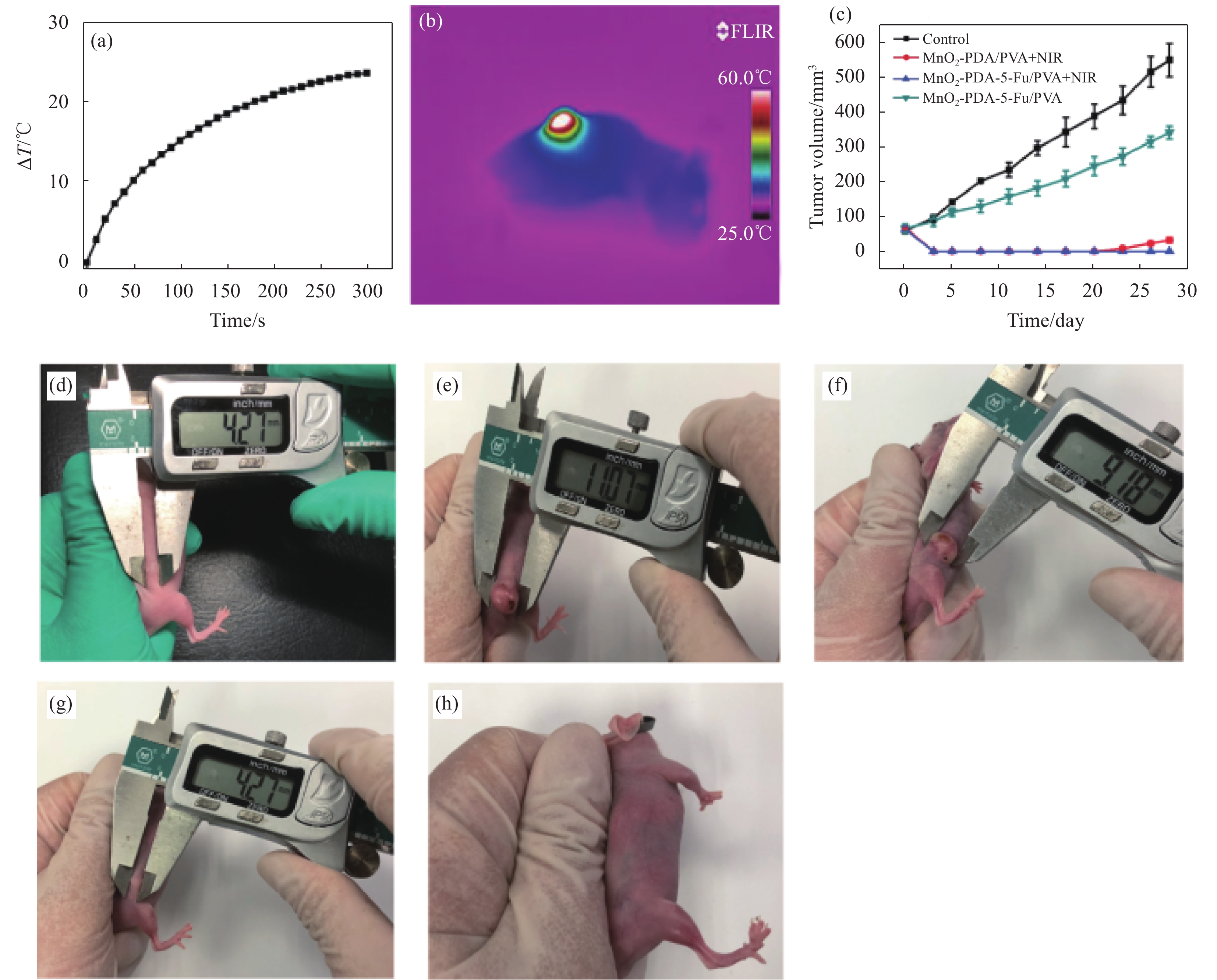
图 8 小鼠光热治疗的温度变化曲线 (a);对应图8(a)小鼠的热成像照片 (b);不同实验组小鼠的肿瘤体积变化曲线 (c);治疗前肿瘤照片 (d);治疗后第28天:对照组 (e)、MnO2-PVA/PDA+NIR (f)、MnO2-PDA-5-FU/PVA (g)和MnO2-PDA-5-FU/PVA+NIR (h) 治疗过程小鼠肿瘤的图片
Figure 8. Temperature curve of mice after the photothermal therapy (a); tumor photothermal imaging corresponding to fig.8(a) (b); tumor volume change curves of mice in different experimental groups (c); tumor photograph before therapy (d); mouse photographs at day 28 after the therapy: Control group (e), MnO2-PVA/PDA+NIR (f), MnO2-PDA-5-FU/PVA (g) and MnO2-PDA-5-FU/PVA+NIR (h)
-
[1] ABAZARI M F, NEJATI F, NASIRI N, et al. Platelet-rich plasma incorporated electrospun PVA-chitosan-HA nano-fibers accelerates osteogenic differentiation and bone reconstruction[J]. Gene,2019,720:144096. doi: 10.1016/j.gene.2019.144096 [2] ZHU H J, CHENG P H, CHEN P, et al. Recent progress in the development of near-infrared organic photothermal and photodynamic nanotherapeutics[J]. Biomaterials Science,2018,6(4):746-765. doi: 10.1039/C7BM01210A [3] NADEM S, ZIYADI H, HEKMATI M, et al. Cross-linked poly(vinyl alcohol) nanofibers as drug carrier of clindamycin[J]. Polymer Bulletin,2019,123:68-82. [4] XIE C C, DING R, WANG X Y, et al. A disulfiram-loaded electrospun poly(vinylidene fluoride) nanofibrous scaffold for cancer treatment[J]. Nanotechnology,2020,31(11):115101. doi: 10.1088/1361-6528/ab5b35 [5] LIU Y Y, XI Y X, ZHAO J L, et al. Preparation of therapeutic-laden konjac hydrogel for tumor combination therapy[J]. Chemical Engineering Journal,2019,375:122048. doi: 10.1016/j.cej.2019.122048 [6] 黄红娜, 张丹参, 张力, 等. 纳米药物载体系统的研究[J]. 河北北方学院学报(医学版), 2010, 27(2):69-71.HUANG Hongna, ZHANG Danshen, ZHANG Li, et al. Study on nanoparticle drug carrier system[J]. Journal of Hebei North University (Medical Edition),2010,27(2):69-71(in Chinese). [7] 金丽霞. 纳米药物载体的研究及临床应用[J]. 中国组织工程研究与临床康复, 2010, 14(8):1429-1432.JIN lixia. Research and clinical application of nano-drug carriers[J]. Journal of Clinical Rehabilitative Tissue En-gineering Research,2010,14(8):1429-1432(in Chinese). [8] LV H, TANG D, SUN Z, et al. Electrospun PCL-based polyurethane/HA microfibers as drug carrier of dexamethasone with enhanced biodegradability and shape memory performances[J]. Colloid and Polymer Science,2020,298(1):103-111. doi: 10.1007/s00396-019-04568-5 [9] YU Y, FENG R, LI J, et al. A hybrid genipin-crosslinked dual-sensitive hydrogel/nanostructured lipid carrier ocular drug delivery platform[J]. Asian Journal of Pharmaceutical Sciences,2019,14(4):423-434. [10] XIAO J, CHENG L, FANG T, et al. Nanoparticle-embedded electrospun fiber–covered stent to assist intraluminal photodynamic treatment of oesophageal cancer[J]. Small,2019,15(49):1904979. doi: 10.1002/smll.201904979 [11] 王东伟, 房宽峻, 刘秀明, 等. 彩色聚合物微球的制备及其在纺织品印染中应用的研究进展[J]. 纺织学报, 2019, 40(3):175-182.WANG Dongwei, FANG Kuanjun, LIU Xiuming, et al. Preparation of color polymer microspheres and research progress thereof in textile dyeing and printing[J]. Journal of Textile Research,2019,40(3):175-182(in Chinese). [12] 王春颖. 功能聚合物微球的制备及性质研究[D]. 长春: 长春理工大学, 2018.WANG Chunying. Preparation and properties of functional polymer microspheres[D]. Changchun: Changchun University of Science and Technology, 2018(in Chinese). [13] 顾炜. 多功能聚合物微球的制备及在癌症诊疗中的应用[D]. 苏州: 苏州大学, 2016.GU Wei. Preparation of multifunctional polymer microspheres and their application in cancer diagnosis and treatment[D]. Suzhou: Soochow University, 2016(in Chinese). [14] ZHU C, YANG H, SHEN L, et al. Microfluidic preparation of PLGA microspheres as cell carriers with sustainable rapa release[J]. Journal of Biomaterials Science Polymer Edition,2019,30(9):1-18. [15] 徐浩, 李娟, 朱晶心, 等. 以电纺丝蛋白纤维为模板的pH响应性介孔SiO2纳米管的制备及药物释放[J]. 复合材料学报, 2020, 37(1):173-181.XU Hao, LI Juan, ZHU Jingxin, et al. Preparation and drug release of pH-responsive mesoporous SiO2 nanotubes by electrospun silk fibroin nanofibers as templates[J]. Acta Materiae Compositae Sinica,2020,37(1):173-181(in Chinese). [16] ADELI H, KHORASANI M T, PARVAZINIA M. Wound dressing based on electrospun PVA/chitosan/starch nanofibrous mats: Fabrication, antibacterial and cytocompatibility evaluation and in vitro healing assay[J]. International Journal of Biological Macromolecules,2019,122:238-254. doi: 10.1016/j.ijbiomac.2018.10.115 [17] DING J X, ZHANG J, LI J N, et al. Electrospun polymer biomaterials[J]. Progress in Polymer Science,2019,90:1-34. doi: 10.1016/j.progpolymsci.2019.01.002 [18] OSANLOO M, ARISH J, SERESHTI H. Developed methods for the preparation of electrospun nanofibers containing plant-derived oil or essential oil: A systematic review[J]. Polymer Bulletin,2020, 77:6085-6104. [19] SAGHAZADEH S, RINOLDI C, SCHOT M, et al. Drug delivery systems and materials for wound healing applications[J]. Advanced Drug Delivery Reviews,2018,127:138-166. doi: 10.1016/j.addr.2018.04.008 [20] 于万永, 赵磊, 王宁, 等. 聚乙烯醇-聚吡咯复合纳米纤维的制备及其导电性能[J]. 复合材料学报, 2018, 35(5):1059-1065.YU Wanyong, ZHAO Lei, WANG Ning, et al. Fabrication and conductive properties of polyvinyl alcohol-polypyrrole composite nanofibers[J]. Acta Materiae Compositae Sinica,2018,35(5):1059-1065(in Chinese). [21] ASADI H, GHAEE A, NOURMOHAMMADI J, et al. Electrospun zein/graphene oxide nanosheet composite nano-fibers with controlled drug release as antibacterial wound dressing[J]. International Journal of Polymeric Materials,2020,69(3):1-13. [22] TAEMEH M A, SHIRAVANDI A, KORAYEM M A, et al. Fabrication challenges and trends in biomedical applications of alginate electrospun nanofibers[J]. Carbohydrate Polymers,2020,228:115419. doi: 10.1016/j.carbpol.2019.115419 [23] XU Y J, ZHAO J L, ZHANG Z L, et al. Preparation of electrospray ALG/PDA-PVP nanocomposites and their application in cancer therapy[J]. Soft Matter,2020,16(1):132-141. doi: 10.1039/C9SM01584A [24] 王栋, 宣丽慧, 李超, 等. 静电纺纤维素纳米晶体/壳聚糖-聚乙烯醇复合纳米纤维的制备与表征[J]. 复合材料学报, 2018, 35(4):964-972.WANG Dong, XUAN Lihui, LI Chao, et al. Preparation and characterization of electrospun cellulose nanocrystals/chitosan-polyvinyl alcohol composite nanofibers[J]. Acta Materiae Compositae Sinica,2018,35(4):964-972(in Chinese). [25] CHEN Q, LUO Y, DU W X, et al. Clearable theranostic platform with a pH-independent chemodynamic therapy enhancement strategy for synergetic photothermal tumor therapy[J]. ACS Applied Materials & Interfaces,2019,11(20):18133-18144. doi: 10.1021/acsami.9b02905 [26] HUANG H, LI K, LIU Q, et al. Dual-response CuS@MnO2 nanoparticles with activatable CT/MR-enhanced in vivo imaging guided photothermal therapy[J]. RSC Advances,2019,9(5):2718-2730. doi: 10.1039/C8RA08637K [27] WANG S, ZHAO J, YANG H, et al. Bottom-up synthesis of WS2 nanosheets with synchronous surface modification for imaging guided tumor regression[J]. Acta Biomaterials,2017,58:442-454. doi: 10.1016/j.actbio.2017.06.014 [28] WU C Y, WANG S G, ZHAO J L, et al. Biodegradable Fe(III)@WS2-PVP nanocapsules for redox reaction and TME-enhanced nanocatalytic, photothermal, and chemotherapy[J]. Advanced Functional Materials,2019,29(26):1901722. doi: 10.1002/adfm.201901722 [29] YUE L D, WANG J L, DAI Z C, et al. pH-responsive, self-sacrificial nanotheranostic agent for potential in vivo and in vitro dual modal MRI/CT imaging, real-time, and in situ monitoring of cancer therapy[J]. Bioconjugate Chemistry,2017,28(2):400-409. doi: 10.1021/acs.bioconjchem.6b00562 [30] PACELLI S, RAMPETSREITER K, MODARESI S, et al. Fabrication of a double-cross-linked interpenetrating polymeric network (IPN) hydrogel surface modified with polydopamine to modulate the osteogenic differentiation of adipose-derived stem cells[J]. ACS Applied Materials & Interfaces,2018,10(30):24955-24962. [31] CHEN Y K, ZHAO J L, WANG S G, et al. Photothermal composite nanomaterials for multimodal tumor therapy under MRI guidance[J]. ChemistrySelect,2019,4(37):11156-11164. doi: 10.1002/slct.201903481 [32] BAO Z, LIU X, LIU Y, et al. Near-infrared light-responsive inorganic nanomaterials for photothermal therapy[J]. Asian Journal of Pharmaceutical Sciences,2016,11(3):349-364. doi: 10.1016/j.ajps.2015.11.123 [33] GUAN G Q, WANG X, LI B, et al. “Transformed” Fe3S4 tetragonal nanosheets: A high-efficiency and body-clearable agent for magnetic resonance imaging guided photothermal and chemodynamic synergistic therapy[J]. Nanoscale,2018,10(37):17902-17911. doi: 10.1039/C8NR06507A [34] TIAN Q W, JIANG F R, ZOU R J, et al. Hydrophilic Cu9S5 nanocrystals: A photothermal agent with a 25.7% heat conversion efficiency for photothermal ablation of cancer cells in vivo[J]. ACS Nano,2011,5(12):9761-9771. doi: 10.1021/nn203293t -

 下载:
下载:


 点击查看大图
计量
- 文章访问数: 1897
- HTML全文浏览量: 626
- PDF下载量: 97
- 被引次数: 0
