


Research advances in 3D printed bone tissue engineering scaffolds based on biodegradable polyester/bioceramics
-
摘要: 移植骨植入物是目前治疗骨缺损的公认有效手段之一。生物可降解聚酯/生物陶瓷复合材料结合了生物可降解聚酯的良好力学性能、可降解性能和生物陶瓷的成骨活性,为骨植入物材料提供了新的选择。骨组织工程通过模拟骨骼微环境,加速骨缺损修复。将生物可降解聚酯/生物陶瓷复合材料制备成骨组织工程支架,能进一步加快骨修复进程。3D打印技术的引入能使生物可降解聚酯/生物陶瓷骨组织工程支架的制备过程精确、可重复且具备高自由度,展现出了良好的发展前景。本文阐述了骨组织工程支架应具备的各项性能,总结了近年来国内外学者对生物可降解聚酯/生物陶瓷骨组织工程支架上述性能的改善策略,并展望未来该研究领域的发展方向。Abstract: Transplantation of bone implants is currently recognized as one of the effective means treating bone defects. Biodegradable polyester/bioceramics composites combine good mechanical and degradable properties of biodegradable polyester with the osteogenic activity of bioceramics, thereby providing a new alternative for bone implant materials. Bone tissue engineering accelerates bone defect repair by simulating the bone microenvironment. The fabrication of biodegradable polyester/bioceramics composites into bone tissue engineering scaffolds can further accelerate the process of bone repair, and the introduction of 3D printing technology enables the preparation of biodegradable polyester/bioceramics bone tissue engineering scaffolds more precise, reproducible, and flexible, which exhibits very promising development. This review presents physical properties of bone tissue engineering scaffolds, summarizes the strategies from domestic and foreign scholars to improve the performance of bone tissue engineering scaffolds based on biodegradable polyester/bioceramics composite in recent years. Besides, the future development perspectives in this field are proposed in the field of research.
-
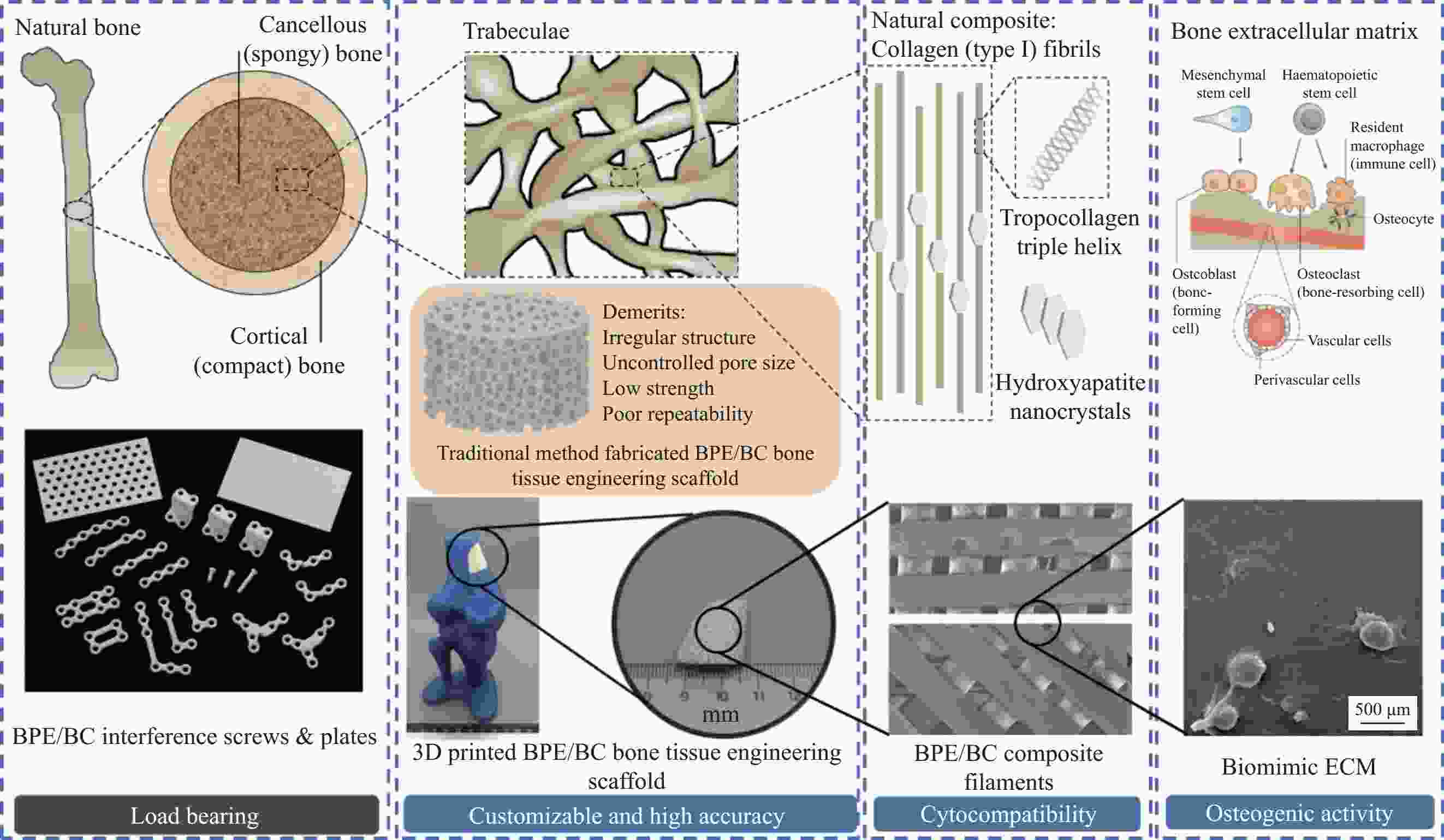
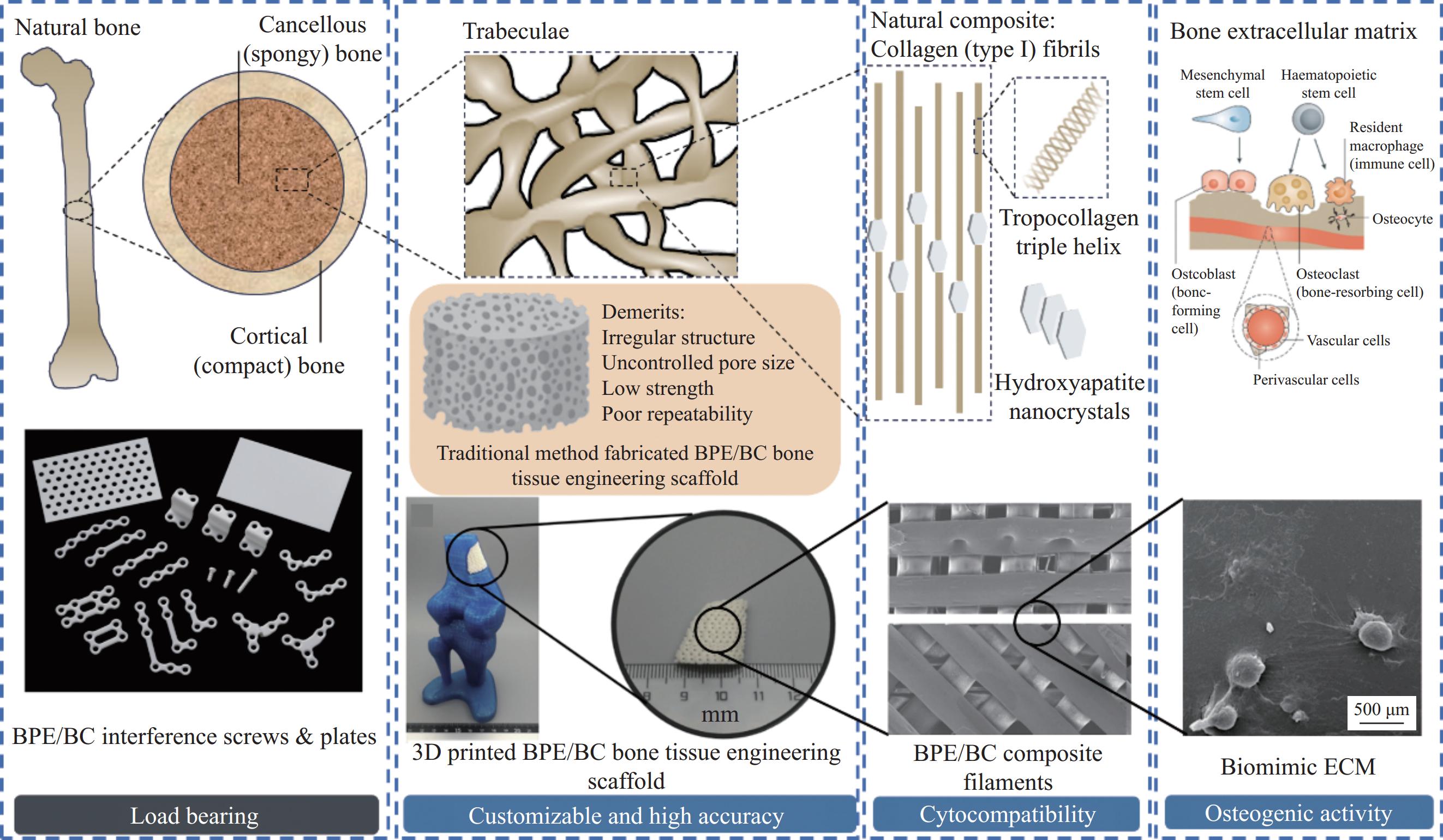
图 1 基于自然骨结构的骨植入物设计
Figure 1. Bone implant design based on natural bone structure
BPE—Biodegradable polyester; BC—Bioceramics; ECM—Extracellular matrix
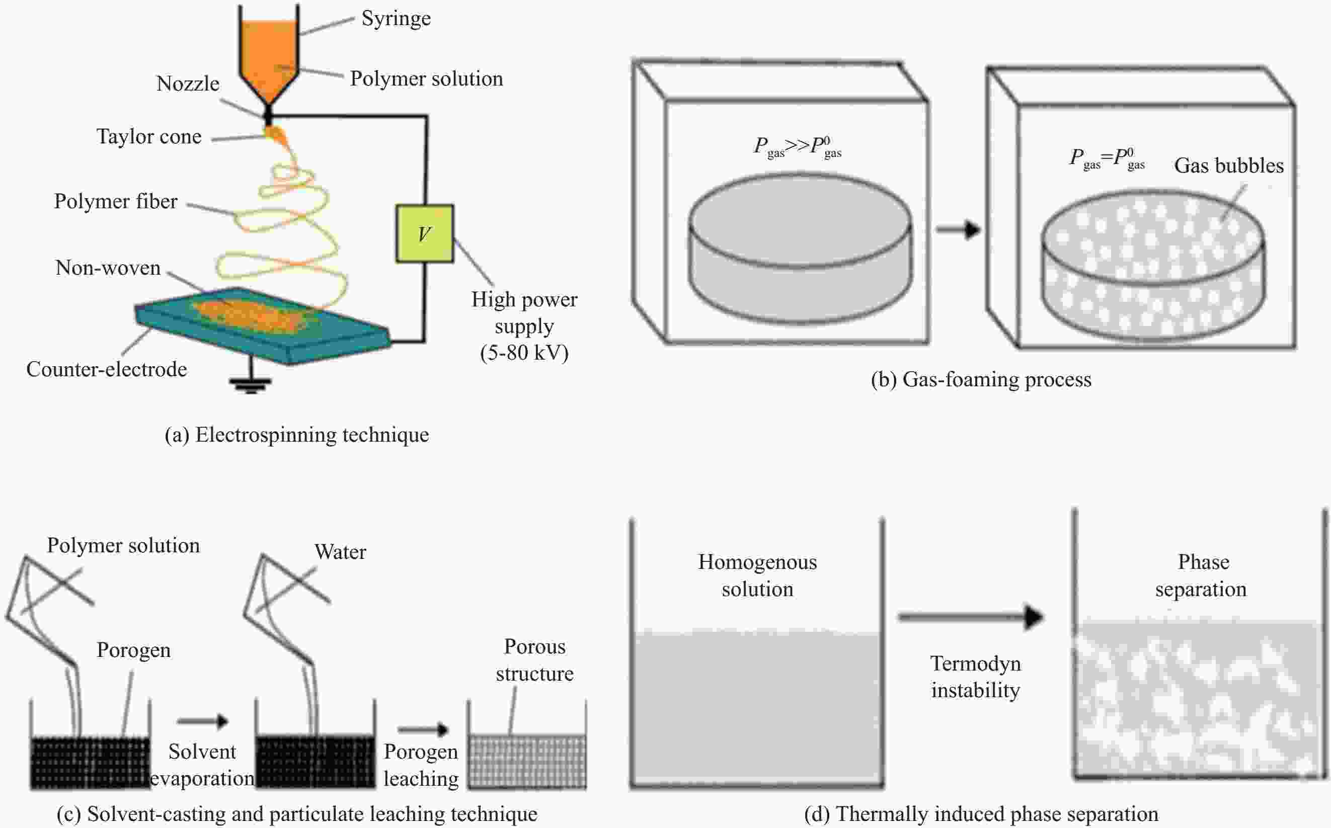
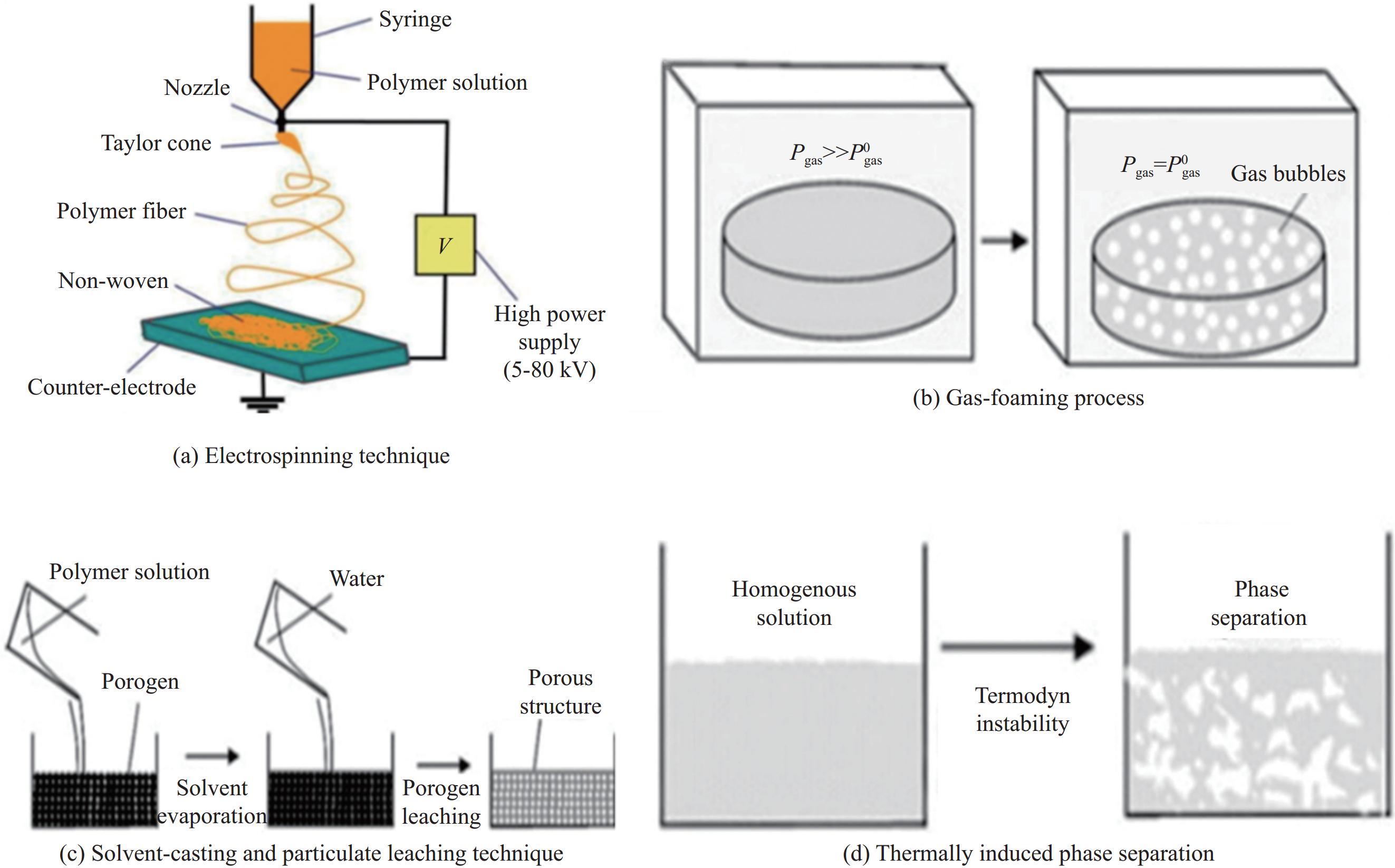
图 2 支架制造技术示意图
Figure 2. Schematic illustrations of classical scaffold fabrication techniques
Pgas—Current gas pressure; Pgas 0 —Equilibrium gas pressure
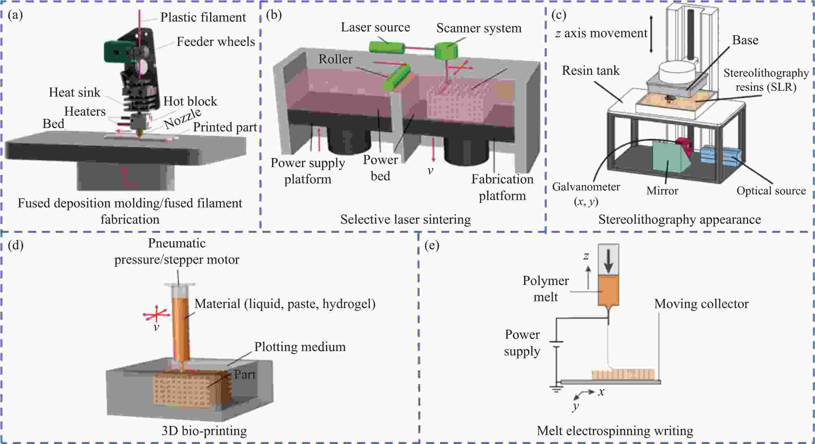
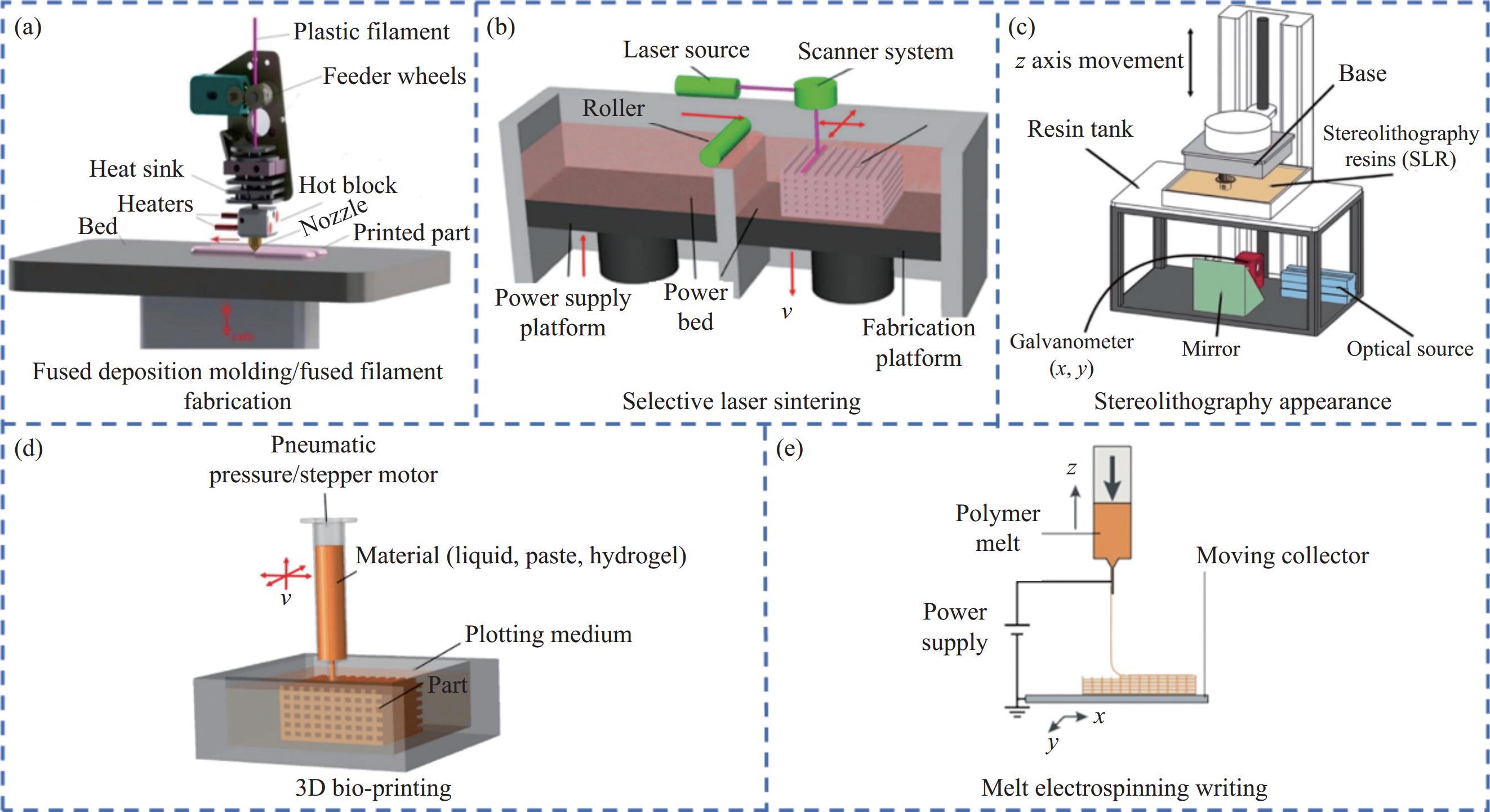
图 3 各种3D打印工艺的示意图:(a) 熔融沉积建模/熔丝制造[17];(b) 扫描激光烧结[19];(c) 立体光刻打印[20];(d) 3D生物打印[21];(e) 熔融电写[22]
Figure 3. A compilation/schematic of various 3D printing processes: (a) Fused deposition molding/fused filament fabrication[17]; (b) Selective laser sintering[19]; (c) Stereolithography[20]; (d) 3D bio-printing[21]; (e) Melt electrospinning writing[22]
v—Vertical
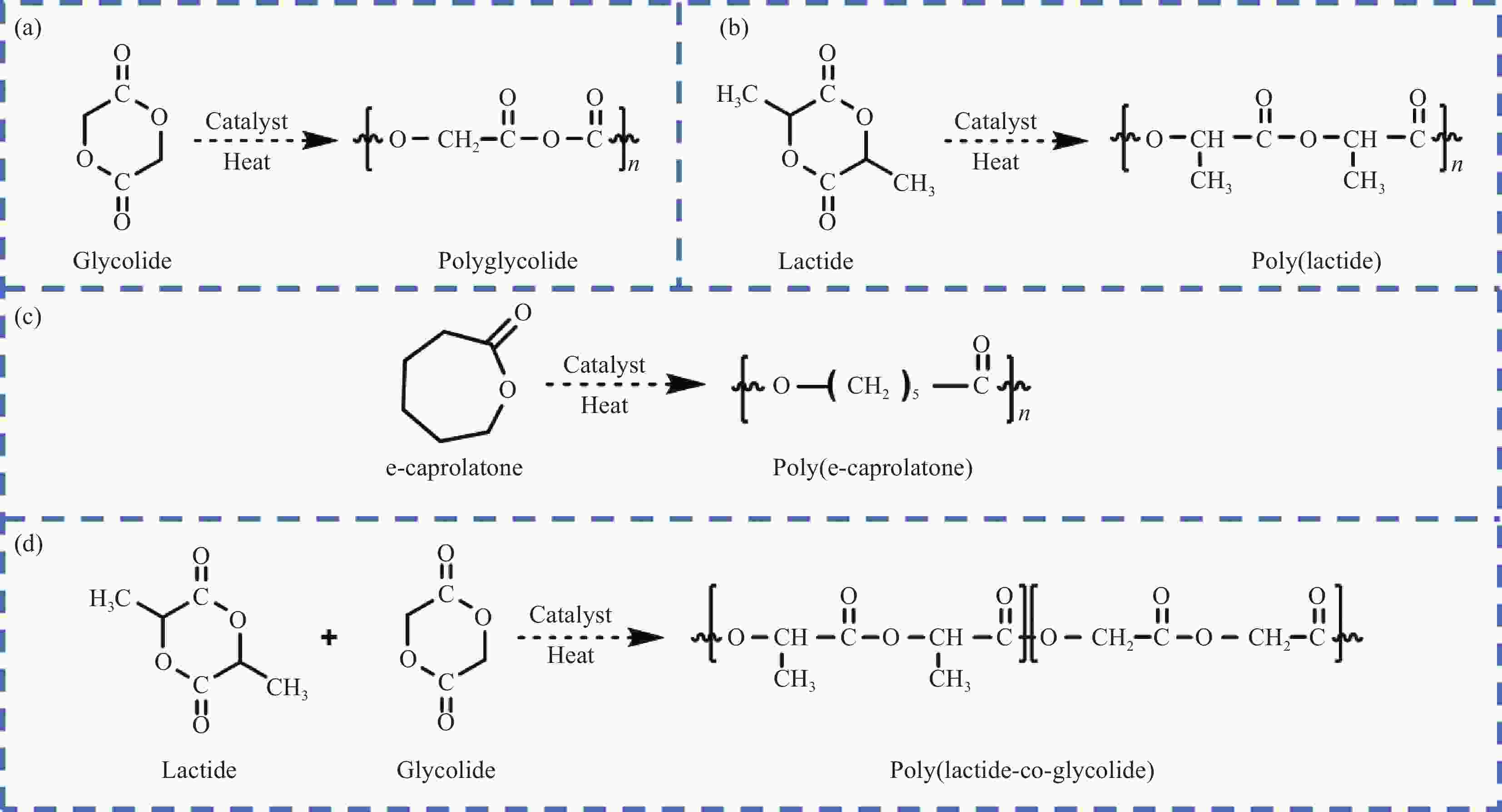
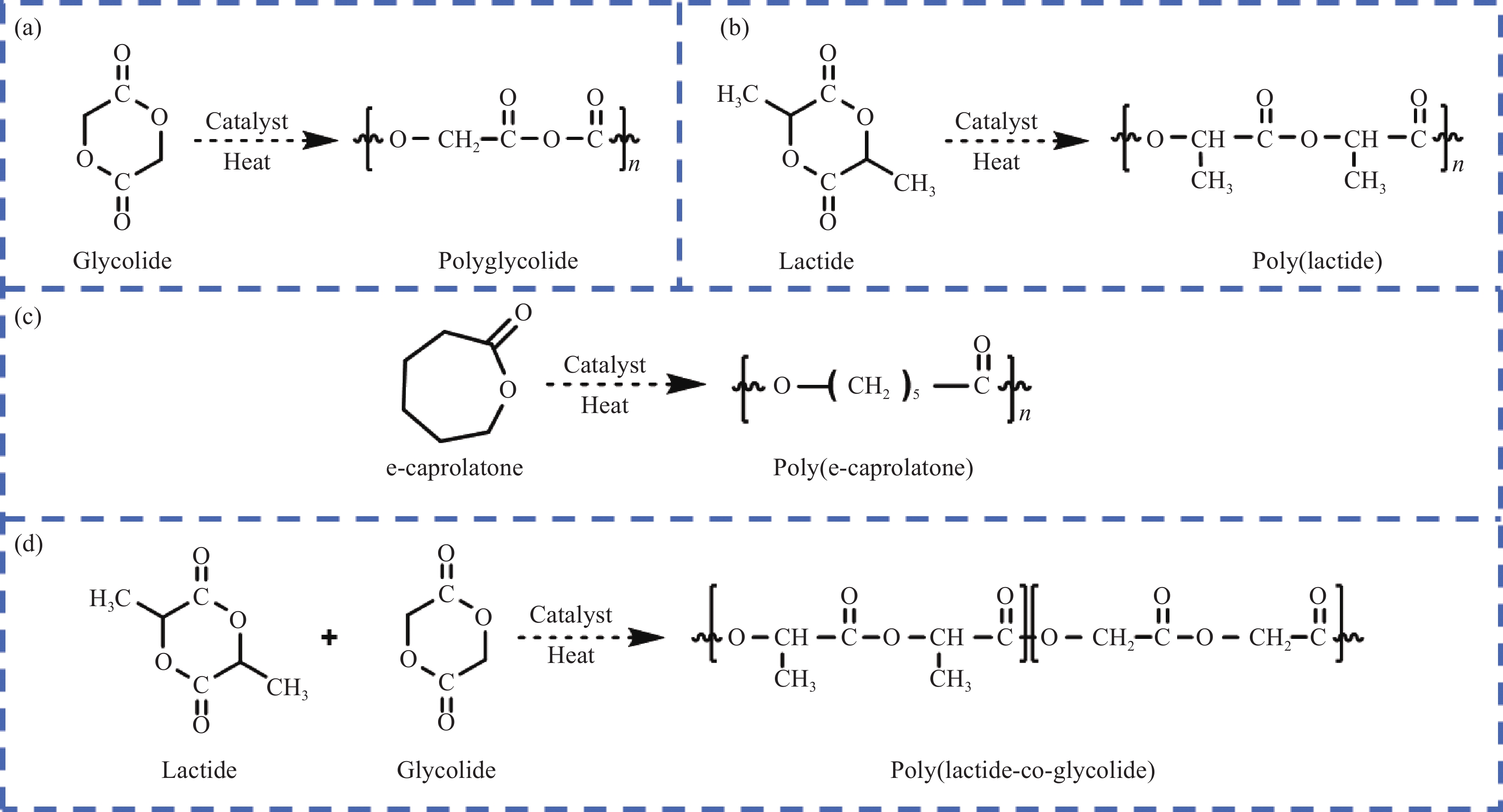
图 4 各类生物可降解聚酯的合成路径:(a) 聚乙醇酸(PGA);(b) 聚乳酸(PLA);(c) 聚己内酯(PCL);(d) 聚乳酸-乙醇酸(PLGA)
Figure 4. Synthetic paths of various biodegradable polyesters: (a) Poly(glycolic acid) (PGA); (b) Poly(lactide acid) (PLA); (c) Poly(e-caprolactone) (PCL); (d) Poly(lactide-co-glycolide) (PLGA)

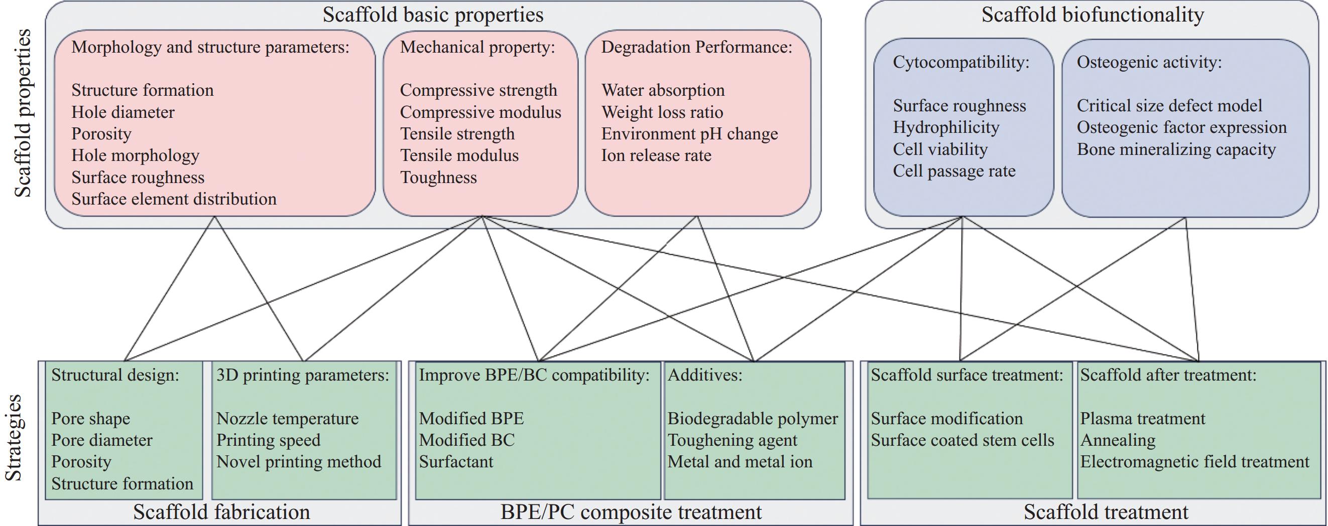
图 5 3D打印生物可降解聚酯/生物陶瓷骨组织工程支架的结构、性能及改进策略
Figure 5. Structure, properties and improvement strategies of 3D-printed biodegradable polyester/bioceramics bone tissue engineering scaffolds
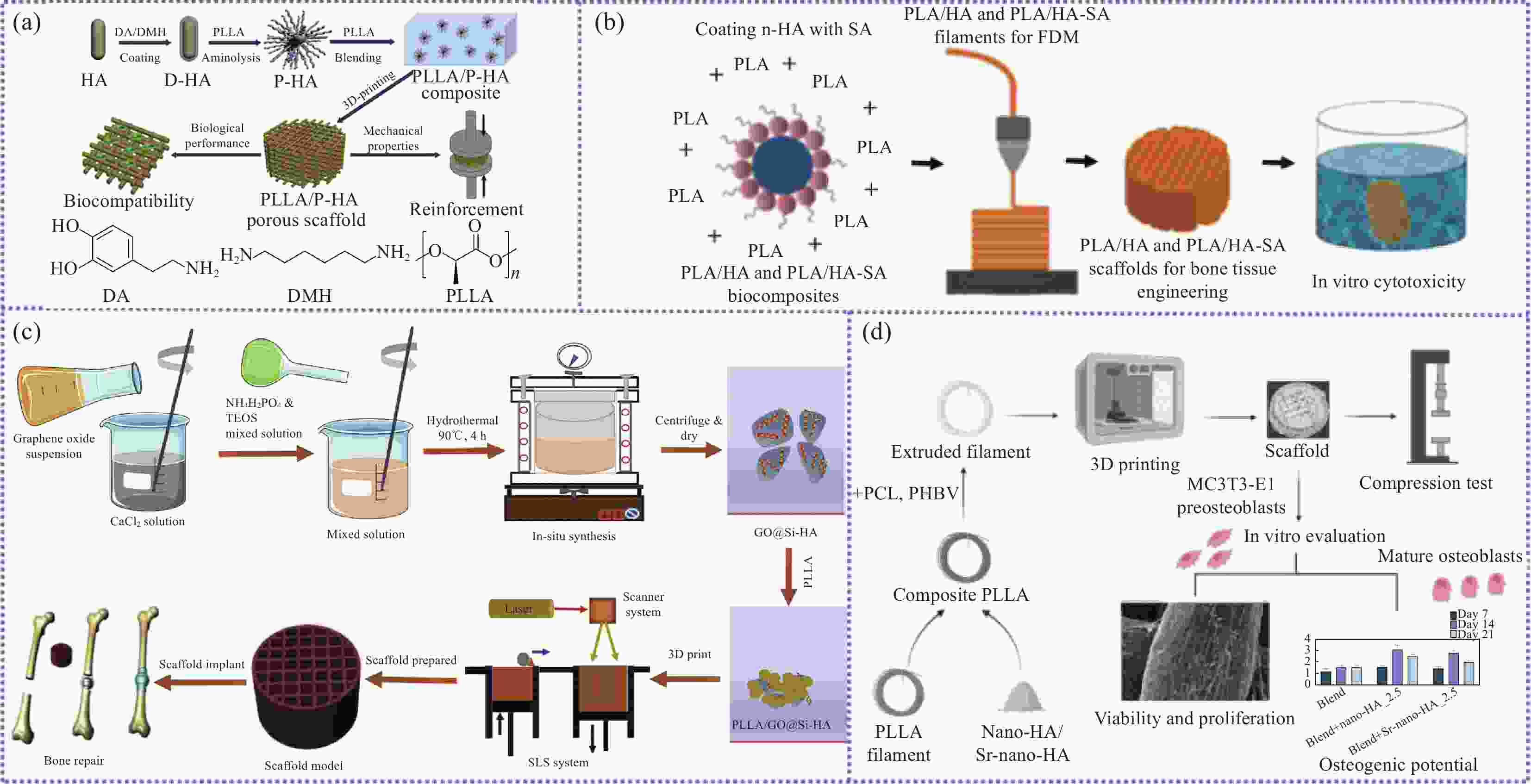
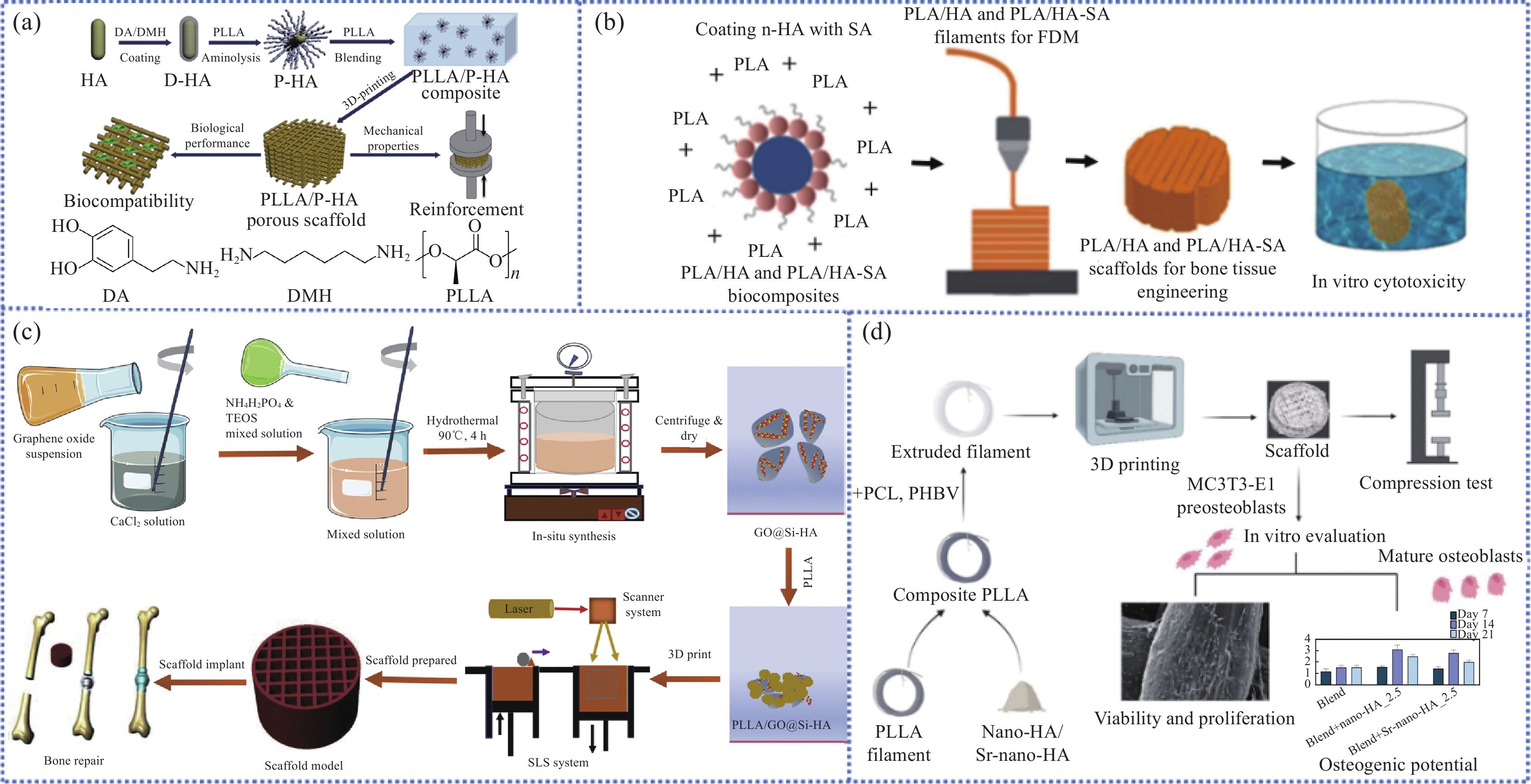
图 6 (a) 左旋聚乳酸(PLLA)/左旋聚乳酸改性羟基磷灰石(P-HA)支架的制备流程[27];(b) 熔融沉积建模(FDM)技术制备PLA-硬脂酸(SA)支架[29];(c) 扫描激光烧结(SLS)技术制备PLLA/氧化石墨烯(GO)@Si-HA复合材料支架[31];(d) PLA/PCL/聚(3-羟基丁酸-3-羟基戊酸) (PHBV)/锶取代纳米羟基磷灰石(Sr-nHA)复合材料支架的制备与表征[32]
Figure 6. (a) Preparation process of poly(L-lactide) (PLLA)/PLLA-modified HA nanoparticles (P-HA) scaffolds[27]; (b) Preparation of PLA-stearic acid (SA) scaffolds by fused deposition modeling (FDM) technique[29]; (c) Preparation of PLLA/graphene oxide (GO)@Si-HA by selected laser sintering (SLS) technique[31]; (d) Preparation and characterization of PLA/PCL/poly(-3-hydroxybutyrate-co-3-hydroxyvalerate) (PHBV)/Sr-nano-hydroxyapatite (Sr-nHA) composite scaffolds[32]
TEOS—Ethyl silicate; D-HA—Poly-dopamine-coated HA nanoparticles; DMH—1, 6-diaminohexane; DA—Dopamine hydrochloride; MC3T3-E1—Mouse embryo osteoblast precursor cells
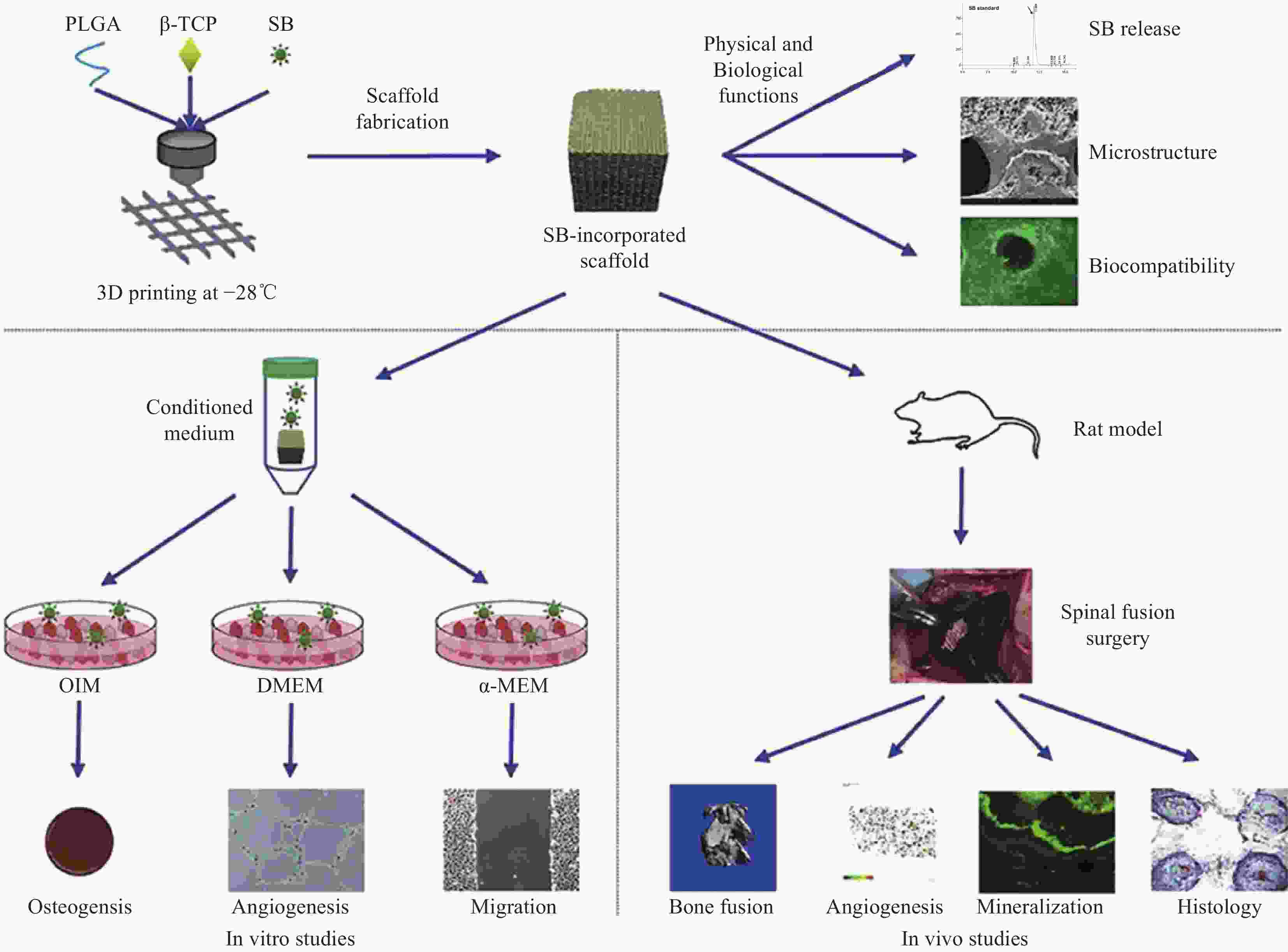
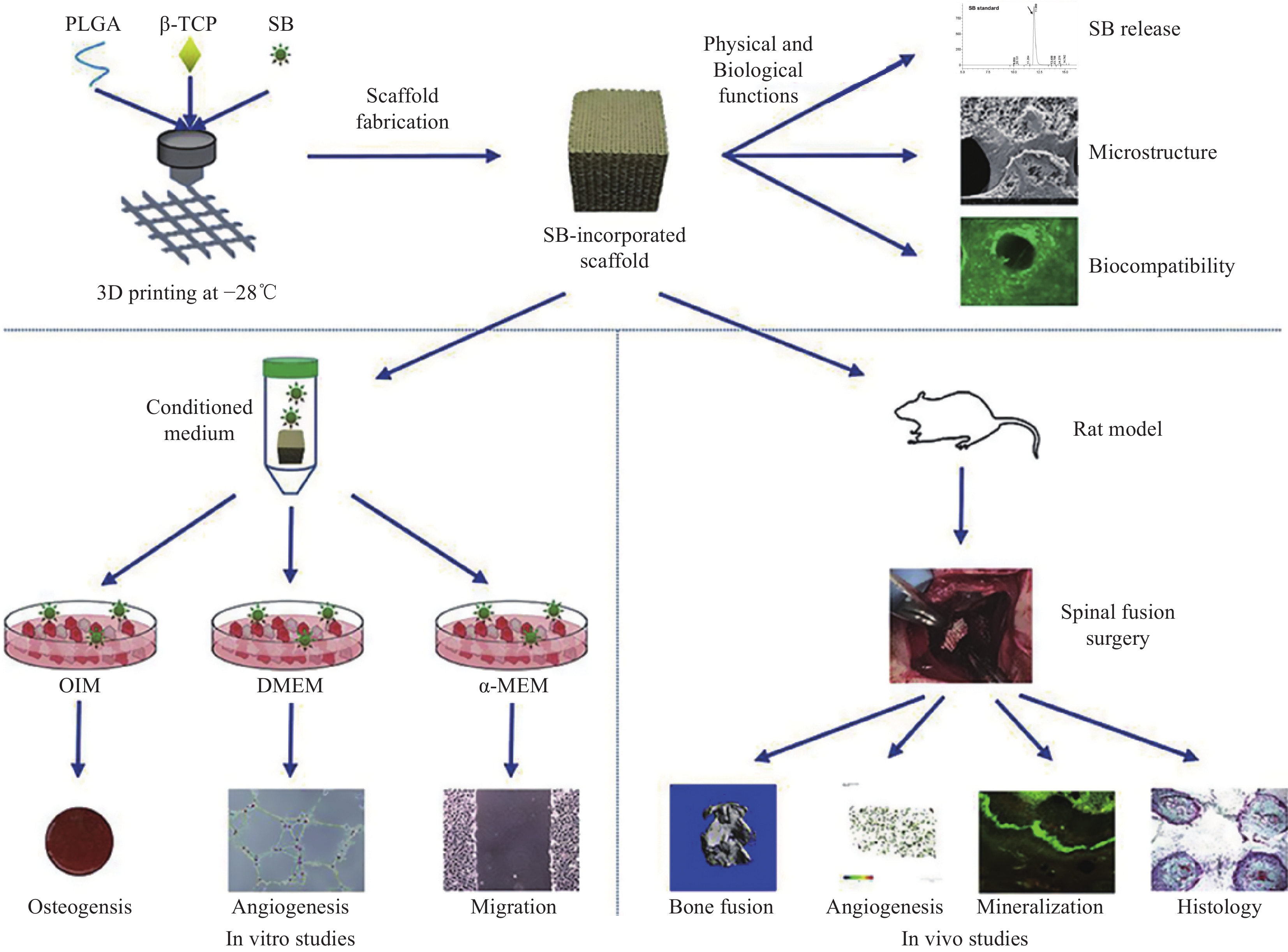
图 7 聚乳酸-乙醇酸(PLGA)/β-磷酸三钙(β-TCP)/丹参酚酸B (SB)支架的制备流程与体内外表征[66]
Figure 7. Preparation process and in vitro and in vivo characterization of polylactic acid-co-polyglycolic acid (PLGA)/β-tricalcium phosphate (β-TCP)/salvianolic acid B (SB) scaffolds[66]
OIM—Osteogenic induction medium; DMEM—Dulbecco's modified eagle medium; α-MEM—Minimum essential medium α
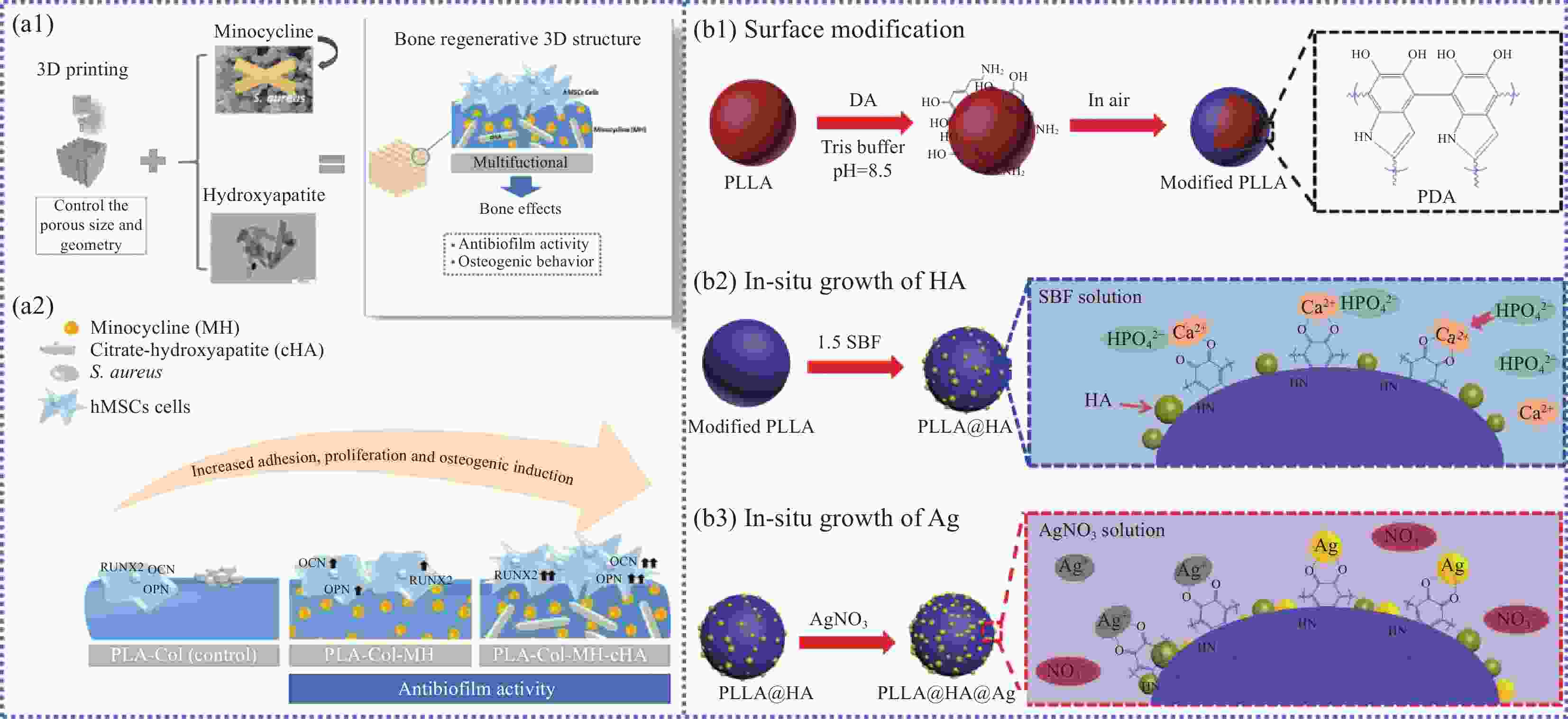
图 8 (a1) PLA-胶原蛋白(Col)-米诺环素(MH)-柠檬酸-羟基磷灰石(cHA)支架的制备流程[46];(a2) PLA-Col、PLA-Col-MH、PLA-Col-MH-cHA支架表面形态[46];((b1)~(b3)) PLLA@HA@Ag颗粒的制备[47]
Figure 8. (a1) Preparation process of PLA-collagen (Col)-minocycline (MH)-citrate-hydroxyapatite (cHA) scaffolds[46]; (a2) Surface morphology of PLA-Col, PLA-Col-MH, and PLA-Col-MH-cHA scaffolds[46]; ((b1)-(b3)) Preparation of PLLA@HA@Ag particles[47]
SBF—Simulated body fluid; hMSCs—Human mesenchymal stem cells; RUNX2—Runt-related transcription factor; OCN—Osteocalcin; OPN—Osteopontin
表 1 典型骨植入物材料性能对比
Table 1. Performance comparison of typical bone implant materials
Types Advantages Drawbacks Natural bone implants Autogenous bone Osteogenic activity, biocompatibility Long surgery time and limited donor bone Homologous bone Wide range of materials, biocompatible Immune response, non-osteogenic activity,
poor mechanical stabilityMetaphyseal bone Low cost, widely available, non-immunogenic Non-osteogenic activity Metals Tantalum, titanium, magnesium alloys Lightweight, high strength, good biocompatibility Complex production process, slow bone
formation, non-degradable, stress maskingBioactive ceramics β-tricalcium phosphate
(β-TCP)Similar composition with bone mineral, good degradability, osteogenic activity, compressive strength Mechanically brittle Bio-glass (BG) Hydroxyapatite (HA) Synthetic polymers Polyetheretherketone (PEEK) High strength, biocompatible, good plasticity Non-degradable Poly(lactic acid) (PLA) Plasticity, biocompatibility,
biodegradabilityInsufficient mechanical properties, non-osteogenic activity Polycaprolactone (PCL) Polyglycolic acid (PGA) Polylactic acid-co-polyglycolic acid (PLGA) Composites PLA/HA Similar to natural bone matrix, good biocompatibility, good osteo-conductivity, low immune-genicity Demanding requirement in manufacture
process 下载: 导出CSV
表 2 传统骨组织工程支架制备方法
Table 2. Preparation methods of bone tissue engineering scaffolds
Preparation methods Preparation principles Advantages Drawbacks Electrospinning [9] Formation of fibers through the influence of an electric field, followed by deposition of the fibers
to produce a highly porous structureLow cost Low connectivity of pores, irregularity, uncon-
trollable pore size, low strength, poor repeatabilityGas foaming[11] Gases grow inside the composite to form a three-dimensional porous polymer structure High porosity, small pore size, no use of organic solvents Solvent casting/particle leaching[12] Lyophilization to remove solvents from the polymer and filtration to obtain a porous structure Simple operation, controlled pore size
and porosityThermally induced phase separation[13] The polymer is dissolved in the solvent at a higher temperature and the porous structure is formed by lowering the temperature until the composite freezes inducing phase separation Good mechanical properties
下载: 导出CSV
表 3 3D打印骨组织工程支架制备方法
Table 3. Preparation methods of 3D printing bone tissue engineering scaffold
3D printing type Working principle Advantage Drawback Fused deposition modeling/fused filament fabrication (FDM/FFF)[17] The polymer in its molten state is prepared as a filament and printed through the spinneret Inexpensive and durable, can be used with a wide range of materials Low resolution, anisotropic, nozzle may be clogged, support required Selected laser sintering
(SLS)[19]Use of laser technology and heat curing Highly durable, easy to remove support material High equipment and material costs, easy to deform prints Stereolithography
(SLA)[20]Laser beam scanning and hardening of
UV-sensitive materialsHigh print accuracy Short laser life and high cost 3D-bioprinting[21] Pressurized syringe extrusion combined with UV curing Highly customizable Uneven micro-morphology Melt electrospinning writing (MEW)[22] Combining the advantages of electrostatic spinning and 3D printing Solvent-free, environmentally friendly and highly efficient Higher cost
下载: 导出CSV
表 4 典型生物可降解聚酯的性能对比
Table 4. Performance comparison of typical biodegradable polyesters
Polyester type Density/(g·cm−3) Tensile strength/MPa Tensile modulus/GPa Tg/℃ Tm/℃ Biodegradation time/month PLA 1.25-1.27 27.6-50.0 1.00-3.45 50-60 — 12-16 PLLA 1.24-1.30 15.5-150 2.7-4.14 55-65 170-200 >24 PDLA 1.21-1.25 21-60 0.35-3.5 45-60 150-162 — PGA 1.50-1.71 60-99.7 6.0-7.0 35-45 220-233 6-12 PCL 1.11-1.14 20.7-42 0.21-0.44 −60-−65 58-65 >24 Notes: PLLA—Poly(L-lactide); PDLA—Poly(D-lactide); Tg—Glass transition temperature; Tm—Melting temperature.
下载: 导出CSV
表 5 文献报道的FDM打印制备的PLA/HA复合材料骨组织工程支架力学性能
Table 5. Mechanical properties of PLA/HA bone tissue engineering scaffolds prepared by FDM printing reported in the literature
Preparation method Compositions Pore size/μm Scaffold mechanical properties Compressive
strength/MPaCompressive
modulus/MPaTensile strength/MPa Tensile modulus/MPa Adding
surfactantsPLA[27] 800 3.13 — — — PLA/PDA-HA[27] 800 4.00 — — — PLLA[28] — 44.02±6.85 — — 43±0.09 PLLA/30HA[28] — 29.68±1.92 — — 45.54±0.11 PLLA/50HA[28] — 14.22±0.63 — — 44.32±0.10 PLA[29] 200 45 — — 56 PLA/SA-HA[29] 200 45 — — 7 Adding other additives PLA[30] 1200 31.34±0.16 598.09±15.24 — — PLA/3WS/3HA[30] 1200 32.15±0.44 506.01±3.52 — — PLA/3WS/8HA[30] 1200 26.83±0.08 721.97±18.27 — — PLA/3WS/15HA[30] 1200 35.72±0.97 883.22±12.32 — — PLA/8WS/3HA[30] 1200 27.58±1.08 723.80±10.23 — — PLA/15WS/3HA[30] 1200 23.36±0.48 680.90±1.93 — — PLLA[31] — 12.1 118 — — PLLA/GO[31] — 18.5 240 — — PLLA/GO@Si-HA[31] — 22.4 280 — — PLLA[32] 800 26.43 — — — PLLA/PCL/PHBV/
2.5Sr-nHA[32]800 32.57 — — — Notes: PDA—Polydopamine; WS—Walnut shell.
下载: 导出CSV
表 6 不同孔隙形状与孔径的3D打印PLA/HA骨组织工程支架的力学性能[33]
Table 6. Mechanical properties of 3D printed PLA/HA bone tissue engineering scaffolds with different pore shapes and pore diameters[33]
Sample Compress strength/MPa Elastic modulus/MPa Poisson's ratio Density/(g·cm−3) Cube-1 5.48 125 0.31 2.9 Cube-2 6.20 136 0.32 2.9 Cylinder-1 6.52 190 0.31 2.9 Cylinder-2 6.70 204 0.33 2.9 Hexgonal-1 7.20 350 0.34 2.9 Hexgonal-2 7.55 410 0.35 2.9
下载: 导出CSV
表 7 文献报道的生物可降解聚酯/生物陶瓷骨组织工程支架性能参数
Table 7. Performance parameters of biodegradable polyester/bioceramic bone tissue engineering scaffolds prepared reported in the literatures
Biomaterial Mixing mass ratio (Polyester :
Bioceramic)Bioceramic particle size Production method Porosity/% Pore size/
μmElastic modulus Compressive strength/MPa Ref. PLA+HA 90:10 50 μm FFF 58 450 — — [75] PLA+CaP coating Uncoated — FDM 49.93±5.28 — (0.512±0.24) GPa 20.50±1.95 [84] 98:2 49.09±3.2 (0.510±0.11) GPa 31.18±4.85 PLA+n-HA 80:20 — FDM 70±2.23 — (10.12±1.24) GPa 31.18±4.86 [81] PLLA+β-TCP 100:0 250 μm 3D bioplotter ~62 100 — 258±102 [85] 90:10 18.22±2.67 70:30 31.18±4.86 PLA+AW 70%AW+30%MD Powder (50:50) 55% (90 μm)
15% (0-53 μm)AW-binder jetting/sintering
PLA-FFFAW disks 41.85%±
0.94%
PLA 60%— — — [86] PLA+HA 85:15 (2.1±0.4) μm MDS 60±1.5 500±20 — — [87] PLA+OCP 40:60 — MEB — 500 — 1 [70] PLA+n-HA 100:0 50-80 nm FDM ~50 — — ~35 [88] 90:10 ~29 80:20 ~28 70:30 ~25 60:40 ~23 50:50 ~17 PLA+n-HA 100:0 (75±20) nm FDM — 300-400 — 35.41±2.07 [89] 50:50 17.80±1.92 PLLA+n-HA 100:0 50-80 nm FDM 60 — (43±0.09) MPa 44.02±6.85 [28] 70:30 (45.54±0.11) MPa 29.68±1.92 50:50 (44.31±0.10) MPa 14.22±0.20 PLA+HA 70:30 50 μm 3D bioplotter 60 500 — — [90] PLA+n-HA 90:10 (63±1.5) nm FDM 26.4 292±1.8 — 23.36±0.48 [91] PLA+HA 85:15 (2.1±1) μm MDS 60 500 — — [82] PLA+HA — — FDM — — — — [82] Notes: AW—Apatite-wollastonite; MD—Maltodexrin; OCP—Octacalcium phosphate; MDS—Mini-deposition system; MEB—Micro-extrusion biopinting.
下载: 导出CSV
表 8 文献报道的生物可降解聚酯/生物陶瓷骨组织工程支架的体内测试方案
Table 8. In vivo testing protocols for biodegradable polyester/bioceramic bone tissue engineering scaffoldsreported in the literatures
Biomaterial groups Animal Sample size Defect CSD Empty control Groups Bone metabolisms substances Sacrifice weeks Main findings Ref. PLA/HA Rat wistar
300 g
4 m/o
1/2 female
1/2 male24 Calvarial bone circular bilateral defect diameter 5.5 mm Yes Yes Empty (n=8)
Bio-oss (n=8)
PLA (n=8)
PLA/HA (n=8)
PLA/HA+DPSC (n=8)
PLA/HA+ECM
(n=8)DPSC
ECM8 w Osteogenic capacity is comparable to
bio-oss.[75] PLA/CaP Rat wistar
300-400 g45 Calvarial bone circular unilateral defect diameter 8 mm Yes No PLA (n=15)
PLA/CaP (n=15)
PLA/CaP+rhBMP-2 (n=15)rhBMP-2 1 m, 3 m and 6 m Promoting new bone formation after 6 months without rhBMP-2. [84] PLA/
n-HARat sprague-dawley
280-320 g weight
12-13 w/o male126 Calvarial bone
circular unilateral defect
diameter 6 mmNo Yes Empty (n=24)
PLA/n-HA (n=24)
PLA/n-HA+EMF
(n=24)
PLA/n-HA+EMF+
BMSCs
(n=24)BMSCs
sinusoidal EMF;
15 Hz, 1 mT
4 h/day4 w and 12 w PLA/HA/BMSCs scaffolds with EMF exposure present the best bone integration among all the groups. [81] PLLA/β-TCP Rat sprague-dawley
320-350 g
8 w/o90 Calvarial bone circular unilateral defect
diameter 5 mmNo No PLLA (n=15)
PLLA/TCP10 (n=15)
PLLA/TCP30 (n=15)
PLLA+
MG-63 (n=15)
PLLA/TCP10+
MG-63 (n=15)
PLLA/TCP30+
MG-63 (n=15)MG-63 4 w, 8 w and 12 w Higher bone regeneration rate in those animals with higher percent of TCP and after adding of MG-63 to the scaffolds. [85] PLA/AW Rat sprague-dawley
350 g
adult
male15 Calvarial bone circular unilateral defect diameter 8 mm Yes No PLA discs (n=3)
AW discs (n=6)
AW/PLA discs (n=6)— 12 w AW/PLA scaffold shows highest formed new bone. [86] PLA/HA Rat sprague-dawley
300-350 g
8 w/o
male32 Calvarial bone
circular unilateral defect
diameter 5 mmNo Yes 3DP PLA/HA (n=8)
β-TCP (n=8)
DBM (n=8)
Black control (n=8)— 4 w and 8 w The percentage of new bone area in 3DP PLA/HA scaffolds was larger than in DBM and control groups but less than β-TCP groups. [87] PLA/OCP Rat
6-8 w/o— Calvarial bone
circular unilateral defect
diameter 5 mmNo Yes — La 8 w The scaffolds enhanced bone defect regeneration in vivo. 0.2La-OCP/PLA scaffolds are significantly more likely to enhance bone defect regeneration in vivo than other groups. [70] PLA/n-HA Rabbit
New Zealand
white
2-3 kg, male18 Femoral Diaphysis
circularNo No PLA (n=9)
PLA/n-HA30%
(n=9)— 4 w, 8 w and 12 w — [88] PLA/n-HA Rabbit
New Zealand
white
2-3 kg, male— Femoral Diaphysis circular unilateral defect
diameter 5 mmNo No — — 1 m, 2 m and 3 m The increasing incorporation of n-HA doesn't affect significantly the overall mechanical strength in a limited range (0%-30%), but it really enhances the osteogenesis in vivo. [89] PLLA/n-HA Rabbit
New Zealand
white
2-3 kg, male9 Diaphysis left
Radius
segmental
unilateral defect
15 mmYes No PLLA (n=3)
PLLA/30%n-HA (n=3)
PLLA/50%n-HA (n=3)— 4 w The new bone growth of the PLLA/50%n-HA composite material is significantly higher than that of the PLA group. [28] PLA/HA Rabbit
New Zealand
white
(2.5±0.25) kg, 6 m/o36 Diaphysis left
Radius
segmental
unilateral defect
15 mmYes No ICBG+IM (n=9)
PLA/HA (n=9)
IM+PLA/HA (n=9)
IM+PLA/HA +eBM (n=9)eBM
ICBG
IMemb4 w, 12 w and 16 w The PLLA/50%n-HA has shown a preferable capability of bone regeneration, compared with the PLLA/30%n-HA specimens than the PLLA ones. [90] PLA/n-HA Rabbit
New Zealand
white
(4.2±0.18) kg, 6 m/o3 Distal right Femur circular unilateral defect
diameter 5 mm and depth
10-13 mmNo No PLA/HA (n=3) — 4 w, 8 w and 12 w The IM combined with 3D-printed PLA-HA scaffold and eBM has a bigger efficiency for treatment of large bone defects than the PLA-HA and the IM/PLA-HA groups, and similar to the IM/ICBG group. [91] PLA/HA Rabbit
New Zealand
white
(2.5±0.2) kg, 6 m/o24 Tibial diaphysis Periosteum
cuboid shaped periosteal pockets 10 mm in length and 7.5 mm in diameter— No Experimental group:
PLA/HA+BMSCs with blood vessel
Control group:
PLA/HA+BMSCs without blood vesselBMSCs 4 w and
8 wIn vivo trials confirmed that the printed PLA/n-HA scaffold can enhance osteogenesis and osteo conductivity. It was showing bone formation within the femoral defect at 4, 8 and 12 weeks, and no inflammation signs. [82] Notes: m/o—Months old; w/o—Weeks old; IM—Intramuscular injection; IV—Intravenous injection; BMSCs—Bone marrow mesenchymal stem cells; DBM—Demineralized bone marrow; La—Lanthanum; eBM—Enhanced bone marrow; ICBG—Iliac crest bone graft; IMemb—Induced membranes; w—Weeks; m—Months; DPSC—Dental pulp stem cells; ECM—Extracellular matrix; rhBMP-2—Recombinant human protein; EMF—Electromagneticfields; MG63—Human osteoblast-like cell; 3DP—3D printing; n—Sample size; CSD—Critical-sized defect.
下载: 导出CSV
-
[1] WANG W, YEUNG K W K. Bone grafts and biomaterials substitutes for bone defect repair: A review[J]. Bioactive Materials, 2017, 2(4): 224-247. doi: 10.1016/j.bioactmat.2017.05.007 [2] GIANNOUDIS P V, DINOPOULOS H, TSIRIDIS E. Bone substitutes: An update[J]. Injury, 2005, 36: S20-S27. [3] TAKIZAWA T, NAKAYAMA N, HANIU H, et al. Titanium fiber plates for bone tissue repair[J]. Advanced Materials, 2018, 30(4): 1703608. doi: 10.1002/adma.201703608 [4] DIMITRIOU R, MATALIOTAKIS G I, ANGOULES A G, et al. Complications following autologous bone graft harvesting from the iliac crest and using the RIA: A systematic review[J]. Injury, 2011, 42: S3-S15. [5] BERGSMA E J, ROZEMA F R, BOS R R, et al. Foreign body reactions to resorbable poly(L-lactide) bone plates and screws used for the fixation of unstable zygomatic fractures[J]. Journal of Oral and Maxillofacial Surgery, 1993, 51(6): 666-670. [6] SURMENEVA M A, CHAIKINA M V, ZAIKOVSKIY V, et al. The structure of an RF-magnetron sputter-deposited silicate-containing hydroxyapatite-based coating investigated by high-resolution techniques[J]. Surface & Coatings Technology, 2013, 218: 39-46. [7] LEGEROS R Z. Calcium phosphate materials in restorative dentistry: A review[J]. Advances in Dental Research, 1988, 2(1): 164-180. doi: 10.1177/08959374880020011101 [8] LEBEDEV S M, GEFLE O S, AMITOV E T, et al. Mechanical properties of PLA-based composites for fused deposition modeling technology[J]. The International Journal of Advanced Manufacturing Technology, 2018, 97(1): 511-518. [9] REN X, LIU Q, ZHENG S, et al. Synergistic delivery of bFGF and BMP-2 from poly(L-lactic-co-glycolic acid)/graphene oxide/hydroxyapatite nanofibre scaffolds for bone tissue engineering applications[J]. RSC Advances, 2018, 8(56): 31911-31923. doi: 10.1039/C8RA05250F [10] COSTANTINI M, BARBETTA A. 6-gas foaming technologies for 3D scaffold engineering[M]//DENG Y, KUIPER J. Functional 3D Tissue Engineering Scaffolds. Cambridge: Woodhead Publishing, 2018: 127-149. [11] COOPER A I. Polymer synthesis and processing using supercritical carbon dioxide[J]. Journal of Materials Chemistry, 2000, 10(2): 207-234. doi: 10.1039/a906486i [12] PANKONGADISAK P, JAIKAEW N, KITI K, et al. The potential use of gentamicin sulfate-loaded poly(L-lactic acid)-sericin hybrid scaffolds for bone tissue engineering[J]. Polymer Bulletin, 2019, 76(6): 2867-2885. doi: 10.1007/s00289-018-2520-x [13] BUZAROVSKA A, DINESCU S, CHITOIU L, et al. Porous poly(L-lactic acid) nanocomposite scaffolds with functionalized TiO2 nanoparticles: Properties, cytocompatibility and drug release capability[J]. Journal of Materials Science, 2018, 53(16): 11151-11166. doi: 10.1007/s10853-018-2415-0 [14] PUPPI D, CHIELLINI F, PIRAS A M, et al. Polymeric materials for bone and cartilage repair[J]. Progress in Polymer Science, 2010, 35(4): 403-440. doi: 10.1016/j.progpolymsci.2010.01.006 [15] ZHANG X, CHEN J L, XING F, et al. Three-dimensional printed polylactic acid and hydroxyapatite composite scaffold with urine-derived stem cells as a treatment for bone defects[J]. Journal of Materials Science: Materials in Medicine, 2022, 33(10): 71. doi: 10.1007/s10856-022-06686-z [16] SENATOV F S, NIAZA K V, ZADOROZHNYY M Y, et al. Mechanical properties and shape memory effect of 3D-printed PLA-based porous scaffolds[J]. Journal of the Mechanical Behavior of Biomedical Materials, 2016, 57: 139-148. doi: 10.1016/j.jmbbm.2015.11.036 [17] HEIDARI-RARANI M, RAFIEE-AFARANI M, ZAHEDI A M. Mechanical characterization of FDM 3D printing of continuous carbon fiber reinforced PLA composites[J]. Composites Part B: Engineering, 2019, 175: 107147. doi: 10.1016/j.compositesb.2019.107147 [18] CHINNASAMI H, DEY M K, DEVIREDDY R. Three-dimensional scaffolds for bone tissue engineering[J]. Bioengineering (Basel), 2023, 10(7): 759. doi: 10.3390/bioengineering10070759 [19] SHUAI C, YANG F, SHUAI Y, et al. Silicon dioxide nanoparticles decorated on graphene oxide nanosheets and their application in poly(L-lactic acid) scaffold[J]. Journal of Advanced Research, 2023, 48: 175-190. doi: 10.1016/j.jare.2022.08.017 [20] ZHOU X, ZHOU G, JUNKA R, et al. Fabrication of polylactic acid (PLA)-based porous scaffold through the combination of traditional bio-fabrication and 3D printing technology for bone regeneration[J]. Colloids and Surfaces B: Biointerfaces, 2021, 197: 111420. doi: 10.1016/j.colsurfb.2020.111420 [21] ZHAO D, WANG Y, YU Z, et al. Co-culture bioprinting of tissue-engineered bone-periosteum biphasic complex for repairing critical-sized skull defects in rabbits[J]. International Journal of Bioprinting, 2023, 9: 698. doi: 10.18063/ijb.698 [22] KOONS G L, DIBA M, MIKOS A G. Materials design for bone-tissue engineering[J]. Nature Reviews Materials, 2020, 5(8): 584-603. doi: 10.1038/s41578-020-0204-2 [23] TRIVEDI A K, GUPTA M K, SINGH H. PLA based biocomposites for sustainable products: A review[J]. Advanced Industrial and Engineering Polymer Research, 2023, 6(4): 382-395. doi: 10.1016/j.aiepr.2023.02.002 [24] SMIESZEK A, TOMASZEWSKI K A, KORNICKA K, et al. Metformin promotes osteogenic differentiation of adipose-derived stromal cells and exerts pro-osteogenic effect stimulating bone regeneration[J]. Journal of Clinical Medicine, 2018, 7(12): 482. doi: 10.3390/jcm7120482 [25] CHEN X, FAN H, DENG X, et al. Scaffold structural microenvironmental cues to guide tissue regeneration in bone tissue applications[J]. Nanomaterials (Basel), 2018, 8(11): 960. doi: 10.3390/nano8110960 [26] WUBNEH A, TSEKOURA E K, AYRANCI C, et al. Current state of fabrication technologies and materials for bone tissue engineering[J]. Acta Biomaterialia, 2018, 80: 1-30. doi: 10.1016/j.actbio.2018.09.031 [27] YANG W F, LONG L, WANG R, et al. Surface-modified hydroxyapatite nanoparticle-reinforced polylactides for three-dimensional printed bone tissue engineering scaffolds[J]. Journal of Biomedical Nanotechnology, 2018, 14(2): 294-303. doi: 10.1166/jbn.2018.2495 [28] ZHANG B, WANG L, SONG P, et al. 3D printed bone tissue regenerative PLA/HA scaffolds with comprehensive performance optimizations[J]. Materials & Design, 2021, 201: 109490. [29] CÉ DE ANDRADE J, CABRAL F, CLEMENS F J, et al. Effect of stearic acid on the mechanical and rheological properties of PLA/HA biocomposites[J]. Materials Today Communications, 2023, 35: 106357. doi: 10.1016/j.mtcomm.2023.106357 [30] SONG X, GUAN W, QIN H, et al. Properties of poly(lactic acid)/walnut shell/hydroxyapatite composites prepared with fused deposition modeling[J]. Scientific Reports, 2022, 12(1): 11563. doi: 10.1038/s41598-022-15622-8 [31] WANG G, QIAN G, ZAN J, et al. A co-dispersion nanosystem of graphene oxide@silicon-doped hydroxyapatite to improve scaffold properties[J]. Materials & Design, 2021, 199: 109399. [32] KONTOGIANNI G I, BONATTI A F, DE MARIA C, et al. Promotion of in vitro osteogenic activity by melt extrusion-based PLLA/PCL/PHBV scaffolds enriched with nano-hydroxyapatite and strontium substituted nano-hydroxyapatite[J]. Polymers (Basel), 2023, 15(4): 1052. doi: 10.3390/polym15041052 [33] SAHMANI S, KHANDAN A, SABER-SAMANDARI S, et al. Fabrication and resonance simulation of 3D-printed biocomposite mesoporous implants with different periodic cellular topologies[J]. Bioprinting, 2021, 22: e00138. doi: 10.1016/j.bprint.2021.e00138 [34] PREMPHET P, LEKSAKUL K, BOONYAWAN D, et al. Process parameters optimization and mechanical properties of 3D PLA/HA printing scaffold[J]. Materials Today: Proceedings, 2023, 4: 124. [35] HWANGBO H, LEE J, KIM G. Mechanically and biologically enhanced 3D-printed HA/PLLA/dECM biocomposites for bone tissue engineering[J]. International Journal of Biological Macromolecules, 2022, 218: 9-21. doi: 10.1016/j.ijbiomac.2022.07.040 [36] PÉREZ-DAVILA S, GARRIDO-GULÍAS N, GONZÁLEZ-RODRÍGUEZ L, et al. Physicochemical properties of 3D-printed polylactic acid/hydroxyapatite scaffolds[J]. Polymers (Basel), 2023, 15(13): 2849. doi: 10.3390/polym15132849 [37] FU Z, CUI J, ZHAO B, et al. An overview of polyester/hydroxyapatite composites for bone tissue repairing[J]. Journal of Orthopaedic Translation, 2021, 28: 118-130. doi: 10.1016/j.jot.2021.02.005 [38] BACKES E H, FERNANDES E M, DIOGO G S, et al. Engineering 3D printed bioactive composite scaffolds based on the combination of aliphatic polyester and calcium phosphates for bone tissue regeneration[J]. Materials Science and Engineering: C, 2021, 122: 111928. doi: 10.1016/j.msec.2021.111928 [39] SHUAI C J, YANG W J, FENG P, et al. Accelerated degradation of HAP/PLLA bone scaffold by PGA blending facilitates bioactivity and osteoconductivity[J]. Bioactive Materials, 2021, 6(2): 490-502. doi: 10.1016/j.bioactmat.2020.09.001 [40] FENG P, PENG S, SHUAI C, et al. In situ generation of hydroxyapatite on biopolymer particles for fabrication of bone scaffolds owning bioactivity[J]. ACS Applied Materials & Interfaces, 2020, 12(41): 46743-46755. [41] BAHRAMINASAB M, DOOSTMOHAMMADI N, TALEBI A, et al. 3D printed polylactic acid/gelatin-nano-hydroxyapatite/platelet-rich plasma scaffold for critical-sized skull defect regeneration[J]. BioMedical Engineering OnLine, 2022, 21(1): 86. doi: 10.1186/s12938-022-01056-w [42] NARAYANAN G, VERNEKAR V N, KUYINU E L, et al. Poly (lactic acid)-based biomaterials for orthopaedic regenerative engineering[J]. Advanced Drug Delivery Reviews, 2016, 107: 247-276. doi: 10.1016/j.addr.2016.04.015 [43] CHIA H N, WU B M. Recent advances in 3D printing of biomaterials[J]. Journal of Biological Engineering, 2015, 9: 4. doi: 10.1186/s13036-015-0001-4 [44] HARRIS J S, BEMENDERFER T B, WESSEL A R, et al. A review of mouse critical size defect models in weight bearing bones[J]. Bone, 2013, 55(1): 241-247. doi: 10.1016/j.bone.2013.02.002 [45] WEI L, WU S, KUSS M, et al. 3D printing of silk fibroin-based hybrid scaffold treated with platelet rich plasma for bone tissue engineering[J]. Bioactive Materials, 2019, 4: 256-260. doi: 10.1016/j.bioactmat.2019.09.001 [46] MARTIN V, RIBEIRO I A, ALVES M M, et al. Engineering a multifunctional 3D-printed PLA-collagen-minocycline-nanohydroxyapatite scaffold with combined antimicrobial and osteogenic effects for bone regeneration[J]. Materials Science and Engineering: C, 2019, 101: 15-26. doi: 10.1016/j.msec.2019.03.056 [47] YANG Y, CHENG Y, DENG F, et al. A bifunctional bone scaffold combines osteogenesis and antibacterial activity via in situ grown hydroxyapatite and silver nanoparticles[J]. Bio-Design and Manufacturing, 2021, 4(3): 452-468. doi: 10.1007/s42242-021-00130-x [48] MARYCZ K, SMIESZEK A, TARGONSKA S, et al. Three dimensional (3D) printed polylactic acid with nano-hydroxyapatite doped with europium(III) ions (nHAp/PLLA@Eu3+) composite for osteochondral defect regeneration and theranostics[J]. Materials Science and Engineering: C, 2020, 110: 110634. doi: 10.1016/j.msec.2020.110634 [49] ZHENG H, SUN Z, ZHANG H. Effects of walnut shell powders on the morphology and the thermal and mechanical properties of poly(lactic acid)[J]. Journal of Thermoplastic Composite Materials, 2019, 33(10): 1383-1395. [50] FERREIRA F V, BRITO F S, FRANCESCHI W, et al. Functionalized graphene oxide as reinforcement in epoxy based nanocomposites[J]. Surfaces and Interfaces, 2018, 10: 100-109. doi: 10.1016/j.surfin.2017.12.004 [51] VILA M, GARCÍA A, GIROTTI A, et al. 3D silicon doped hydroxyapatite scaffolds decorated with elastin-like recombinamers for bone regenerative medicine[J]. Acta Biomaterialia, 2016, 45: 349-356. doi: 10.1016/j.actbio.2016.09.016 [52] HOU Y, WANG W, BARTOLO P. Investigation of polycaprolactone for bone tissue engineering scaffolds: In vitro degradation and biological studies[J]. Materials & Design, 2022, 216: 110582. [53] JIAO Z, LUO B, XIANG S, et al. 3D printing of HA/PCL composite tissue engineering scaffolds[J]. Advanced Industrial and Engineering Polymer Research, 2019, 2(4): 196-202. doi: 10.1016/j.aiepr.2019.09.003 [54] JOHN J, DEVJANI D, ALI S, et al. Optimization of 3D printed polylactic acid structures with different infill patterns using Taguchi-grey relational analysis[J]. Advanced Industrial and Engineering Polymer Research, 2023, 6(1): 62-78. doi: 10.1016/j.aiepr.2022.06.002 [55] ESHRAGHI S, DAS S. Micromechanical finite-element modeling and experimental characterization of the compressive mechanical properties of polycaprolactone-hydroxyapatite composite scaffolds prepared by selective laser sintering for bone tissue engineering[J]. Acta Biomaterialia, 2012, 8(8): 3138-3143. doi: 10.1016/j.actbio.2012.04.022 [56] OSTAFINSKA A, FORTELNÝ I, HODAN J, et al. Strong synergistic effects in PLA/PCL blends: Impact of PLA matrix viscosity[J]. Journal of the Mechanical Behavior of Biomedical Materials, 2017, 69: 229-241. doi: 10.1016/j.jmbbm.2017.01.015 [57] OLEWNIK-KRUSZKOWSKA E, KASPERSKA P, KOTER I. Effect of poly(ε-caprolactone) as plasticizer on the properties of composites based on polylactide during hydrolytic degradation[J]. Reactive and Functional Polymers, 2016, 103: 99-107. doi: 10.1016/j.reactfunctpolym.2016.03.026 [58] ÅKERLUND E, DIEZ-ESCUDERO A, GRZESZCZAK A, et al. The effect of PCL addition on 3D-printable PLA/HA composite filaments for the treatment of bone defects[J]. Polymers (Basel), 2022, 14(16): 3305. doi: 10.3390/polym14163305 [59] ESPOSITO CORCIONE C, GERVASO F, SCALERA F, et al. Highly loaded hydroxyapatite microsphere/PLA porous scaffolds obtained by fused deposition modelling[J]. Ceramics International, 2019, 45(2): 2803-2810. [60] JOSEPH B, NINAN N, VISALAKSHAN R M, et al. Insights into the biomechanical properties of plasma treated 3D printed PCL scaffolds decorated with gold nanoparticles[J]. Composites Science and Technology, 2021, 202: 108544. doi: 10.1016/j.compscitech.2020.108544 [61] LIU C, YAO W, TIAN M, et al. Mussel-inspired degradable antibacterial polydopamine/silica nanoparticle for rapid hemostasis[J]. Biomaterials, 2018, 179: 83-95. doi: 10.1016/j.biomaterials.2018.06.037 [62] WANG H, YANG J, LIU X, et al. A robust 3D superhydrophobic sponge for in situ continuous oil removing[J]. Journal of Materials Science, 2019, 54(2): 1255-1266. doi: 10.1007/s10853-018-2938-4 [63] DOU Y, HUANG J, XIA X, et al. A hierarchical scaffold with a highly pore-interconnective 3D printed PLGA/n-HA framework and an extracellular matrix like gelatin network filler for bone regeneration[J]. Journal of Materials Chemistry B, 2021, 9(22): 4488-4501. doi: 10.1039/D1TB00662B [64] DAVIDENKO N, SCHUSTER C F, BAX D V, et al. Evaluation of cell binding to collagen and gelatin: A study of the effect of 2D and 3D architecture and surface chemistry[J]. Journal of Materials Science-Materials in Medicine, 2016, 27(10): 148. doi: 10.1007/s10856-016-5763-9 [65] CHENG W X, LIU Y Z, MENG X B, et al. PLGA/β-TCP composite scaffold incorporating cucurbitacin B promotes bone regeneration by inducing angiogenesis[J]. Journal of Orthopaedic Translation, 2021, 31: 41-51. doi: 10.1016/j.jot.2021.10.002 [66] LIN S, CUI L, CHEN G, et al. PLGA/β-TCP composite scaffold incorporating salvianolic acid B promotes bone fusion by angiogenesis and osteogenesis in a rat spinal fusion model[J]. Biomaterials, 2019, 196: 109-121. doi: 10.1016/j.biomaterials.2018.04.004 [67] ZHU W, LIANG S, WANG J, et al. Europium-phenolic network coated BaGdF5 nanocomposites for tri-modal computed tomography/magnetic resonance/luminescence imaging[J]. Journal of Materials Science: Materials in Medicine, 2017, 28(5): 74. doi: 10.1007/s10856-017-5888-5 [68] HE J, ZHANG N, ZHANG J, et al. Migration critically meditates osteoblastic differentiation of bone mesenchymal stem cells through activating canonical Wnt signal pathway[J]. Colloids and Surfaces B: Biointerfaces, 2018, 171: 205-213. doi: 10.1016/j.colsurfb.2018.07.017 [69] WANG W, WEI J, LEI D, et al. 3D printing of lithium osteogenic bioactive composite scaffold for enhanced bone regeneration[J]. Composites Part B: Engineering, 2023, 256: 110641. doi: 10.1016/j.compositesb.2023.110641 [70] XU Z, LIN B, ZHAO C, et al. Lanthanum doped octacalcium phosphate/polylactic acid scaffold fabricated by 3D printing for bone tissue engineering[J]. Journal of Materials Science & Technology, 2022, 118: 229-242. [71] HUANG J, LIU W, LIANG Y, et al. Preparation and biocompatibility of diphasic magnetic nanocomposite scaffold[J]. Materials Science and Engineering: C, 2018, 87: 70-77. doi: 10.1016/j.msec.2018.02.003 [72] PORTER R M, LIU F, PILAPIL C, et al. Osteogenic potential of reamer irrigator aspirator (RIA) aspirate collected from patients undergoing hip arthroplasty[J]. Journal of Orthopaedic Research, 2009, 27(1): 42-49. doi: 10.1002/jor.20715 [73] LIU Z, CHU W, ZHANG L, et al. The effect of enhanced bone marrow in conjunction with 3D-printed PLA-HA in the repair of critical-sized bone defects in a rabbit model[J]. Annals of Translational Medicine, 2021, 9(14): 1134. doi: 10.21037/atm-20-8198 [74] PADUANO F, MARRELLI M, ALOM N, et al. Decellularized bone extracellular matrix and human dental pulp stem cells as a construct for bone regeneration[J]. Journal of Biomaterials Science, Polymer Edition, 2017, 28(8): 730-748. doi: 10.1080/09205063.2017.1301770 [75] GENDVILIENE I, SIMOLIUNAS E, ALKSNE M, et al. Effect of extracellular matrix and dental pulp stem cells on bone regeneration with 3D printed PLA/HA composite scaffolds[J]. European Cells and Materials, 2021, 41: 204-215. doi: 10.22203/eCM.v041a15 [76] ZHANG D, WEI G, LI P, et al. Urine-derived stem cells: A novel and versatile progenitor source for cell-based therapy and regenerative medicine[J]. Genes & Diseases, 2014, 1(1): 8-17. [77] BODIN A, BHARADWAJ S, WU S, et al. Tissue-engineered conduit using urine-derived stem cells seeded bacterial cellulose polymer in urinary reconstruction and diversion[J]. Biomaterials, 2010, 31(34): 8889-8901. doi: 10.1016/j.biomaterials.2010.07.108 [78] LIAO H T, LEE M Y, TSAI W W, et al. Osteogenesis of adipose-derived stem cells on polycaprolactone-β-tricalcium phosphate scaffold fabricated via selective laser sintering and surface coating with collagen type I[J]. Journal of Tissue Engineering and Regenerative Medicine, 2016, 10(10): E337-E353. doi: 10.1002/term.1811 [79] ZHENG C, ATTARILAR S, LI K, et al. 3D-printed HA15-loaded β-tricalcium phosphate/poly (lactic-co-glycolic acid) bone tissue scaffold promotes bone regeneration in rabbit radial defects[J]. International Journal of Bioprinting, 2021, 7(1): 317. [80] SONG M, ZHAO D, WEI S, et al. The effect of electromagnetic fields on the proliferation and the osteogenic or adipogenic differentiation of mesenchymal stem cells modulated by dexamethasone[J]. Bioelectromagnetics, 2014, 35(7): 479-490. doi: 10.1002/bem.21867 [81] TU C, CHEN J, HUANG C, et al. Effects of electromagnetic fields treatment on rat critical-sized calvarial defects with a 3D-printed composite scaffold[J]. Stem Cell Research and Therapy, 2020, 11(1): 433. doi: 10.1186/s13287-020-01954-7 [82] ZHANG H, MAO X, ZHAO D, et al. Three dimensional printed polylactic acid-hydroxyapatite composite scaffolds for prefabricating vascularized tissue engineered bone: An in vivo bioreactor model[J]. Scientific Reports, 2017, 7(1): 15255. doi: 10.1038/s41598-017-14923-7 [83] LIU Y, MÖLLER B, WILTFANG J, et al. Tissue engineering of a vascularized bone graft of critical size with an osteogenic and angiogenic factor-based in vivo bioreactor[J]. Tissue Engineering Part A, 2014, 20(23-24): 3189-3197. doi: 10.1089/ten.tea.2013.0653 [84] MAIA-PINTO M O C, BROCHADO A C B, TEIXEIRA B N, et al. Biomimetic mineralization on 3D printed PLA scaffolds: On the response of human primary osteoblasts spheroids and in vivo implantation[J]. Polymers, 2021, 13(1): 74. [85] KWON D Y, PARK J H, JANG S H, et al. Bone regeneration by means of a three-dimensional printed scaffold in a rat cranial defect[J]. Journal of Tissue Engineering and Regenerative Medicine, 2018, 12(2): 516-528. doi: 10.1002/term.2532 [86] TCACENCU I, RODRIGUES N, ALHARBI N, et al. Osseointegration of porous apatite-wollastonite and poly(lactic acid) composite structures created using 3D printing techniques[J]. Materials Science and Engineering: C, 2018, 90: 1-7. doi: 10.1016/j.msec.2018.04.022 [87] ZHANG H, MAO X, DU Z, et al. Three dimensional printed macroporous polylactic acid/hydroxyapatite composite scaffolds for promoting bone formation in a critical-size rat calvarial defect model[J]. Science and Technology of Advanced Materials, 2016, 17(1): 136-148. doi: 10.1080/14686996.2016.1145532 [88] WANG W, ZHANG B, LI M, et al. 3D printing of PLA/n-HA composite scaffolds with customized mechanical properties and biological functions for bone tissue engineering[J]. Composites Part B: Engineering, 2021, 224: 109192. doi: 10.1016/j.compositesb.2021.109192 [89] WANG W, ZHANG B, ZHAO L, et al. Fabrication and properties of PLA/nano-HA composite scaffolds with balanced mechanical properties and biological functions for bone tissue engineering application[J]. Nanotechnology Reviews, 2021, 10(1): 1359-1373. doi: 10.1515/ntrev-2021-0083 [90] LIU Z, GE Y, ZHANG L, et al. The effect of induced membranes combined with enhanced bone marrow and 3D PLA-HA on repairing long bone defects in vivo[J]. Journal of Tissue Engineering and Regenerative Medicine, 2020, 14(10): 1403-1414. doi: 10.1002/term.3106 [91] CHEN X, GAO C, JIANG J, et al. 3D printed porous PLA/nHA composite scaffolds with enhanced osteogenesis and osteoconductivity in vivo for bone regeneration[J]. Biomedical Materials, 2019, 14(6): 065003. doi: 10.1088/1748-605X/ab388d [92] FERREIRA M, RZHEPISHEVSKA O, GRENHO L, et al. Levofloxacin-loaded bone cement delivery system: Highly effective against intracellular bacteria and Staphylococcus aureus biofilms[J]. International Journal of Pharmaceutics, 2017, 532(1): 241-248. doi: 10.1016/j.ijpharm.2017.08.089 [93] SILVA T, GRENHO L, BARROS J, et al. A minocycline-releasing PMMA system as a space maintainer for staged bone reconstructions-in vitro antibacterial, cytocompatibility and anti-inflammatory characterization[J]. Biomedical Materials, 2017, 12(3): 035009. doi: 10.1088/1748-605X/aa68b8 [94] LI X, WANG Y, WANG Z, et al. Composite PLA/PEG/nHA/dexamethasone scaffold prepared by 3D printing for bone regeneration[J]. Macromolecular Bioscience, 2018, 18(6): e1800068. doi: 10.1002/mabi.201800068 [95] WANG Y, YAN L, CHENG R, et al. Multifunctional HA/Cu nano-coatings on titanium using PPy coordination and doping via pulse electrochemical polymerization[J]. Biomaterials Science, 2018, 6(3): 575-585. doi: 10.1039/C7BM01104K [96] RÊGO D F, ELIAS S T, AMATO A A, et al. Anti-tumor effects of metformin on head and neck carcinoma cell lines: A systematic review[J]. Oncology Letters, 2017, 13(2): 554-566. doi: 10.3892/ol.2016.5526 [97] TAN W, GAO C, FENG P, et al. Dual-functional scaffolds of poly(L-lactic acid)/nanohydroxyapatite encapsulated with metformin: Simultaneous enhancement of bone repair and bone tumor inhibition[J]. Materials Science and Engineering: C, 2021, 120: 111592. doi: 10.1016/j.msec.2020.111592 [98] SHI M, XIA L, CHEN Z, et al. Europium-doped mesoporous silica nanosphere as an immune-modulating osteogenesis/angiogenesis agent[J]. Biomaterials, 2017, 144: 176-187. doi: 10.1016/j.biomaterials.2017.08.027 -

 下载:
下载:


 点击查看大图
计量
- 文章访问数: 372
- HTML全文浏览量: 137
- PDF下载量: 50
- 被引次数: 0
