


Research progress of biobased antibacterial hydrogels
-
摘要: 致病微生物所引起的感染一直以来都威胁着全世界人类的健康,抗菌材料在某种情况下可以被视为抗生素的替代品,其中抗菌水凝胶就是一类重要的高分子抗菌材料。生物基抗菌水凝胶根据基体种类不同主要分为壳聚糖基、纤维素基、淀粉基、海藻酸钠基、蛋白质基等,这些基质在自然界具有高丰度,生物相容性及生物可降解性的优点,是制备抗菌水凝胶的理想材料。本文综述了近年来生物基抗菌水凝胶的发展现状及应用领域,着重从生物基抗菌水凝胶的种类、制备和应用3个方面来阐述,最后对生物基抗菌水凝胶所面临的挑战及未来的发展趋势进行了总结和展望。Abstract: Infections caused by pathogenic microorganisms have long been a threat to human health around the world. The development of antibacterial biomaterials can be regarded as a substitute for antibiotics in some cases, among which antibacterial hydrogels are an important class of macromolecular antibacterial agents. Bio-based antibacterial hydrogels can be divided into chitosan, cellulose, starch, sodium alginate and protein-based antibacterial hydrogels according to different matrix types. These substrates have high abundance, good biocompatibility and biodegradability in nature, and are ideal materials for preparing antibacterial hydrogels. In this paper, the development status and application fields of bio-based antibacterial hydrogels in recent years were reviewed, mainly from the types, preparation and application of bio-based antibacterial hydrogels. Finally, the challenges faced by bio-based antibacterial hydrogels and the future development trend were summarized and prospeced.
-

图 1 (a) 壳聚糖(CS)-Ag+/NH3水凝胶机制示意图[17];(b)对照组与CS-Ag+/NH3抗菌活性对比[17];(c) 羧甲基壳聚糖/氧化右旋糖酐/γ-聚谷氨酸(COP)水凝胶交联机制及伤口涂覆示意图[20];(d) COP水凝胶降解示意图[20];(e) 水凝胶抗菌活性及促愈合示意图[20]
Figure 1. (a) Schematic diagram of chitosan (CS)-Ag+/NH3 hydrogel mechanism[17]; (b) Comparison of antibacterial activity between the control group and CS-Ag+/NH3[17]; (c) Schematic diagram of CMCS/OD/γ-PGA (COP) hydrogel crosslinking mechanism and wound coating[20]; (d) Schematic diagram of COP hydrogel degradation[20]; (e) Schematic diagram of hydrogel antibacterial activity and promoting healing[20]
CMCS—carboxymethyl chitosan; γ-PGA—γ-polyglutamic acid; OD—Oxidizing dextran; EDC—1-(3-dimethylaminopropyl)-3-ethylcarbodiimide hydrochloride; NHS—N-Hydroxy succinimide; CO—CMCS/OD; CP—CMCS/γ-PGA; CTS—chitosan;CTS0 —Samples without AgNO3; CTS13—Sample with optimal AgNO3 addition amount

图 2 (a) 羧甲基纤维素(CMC)基水凝胶制备示意图[23];(b) 纤维素和季铵化纤维素(QC)在碱水溶液中与环氧氯丙烷(ECH)交联示意图[24];(c) 细菌纤维素(BC)和3-氨基丙基三乙氧基硅烷(APTES)化学改性反应示意图[26];(d) 菌落计数法检测样品对不同细菌抑制作用[26]
Figure 2. (a) Diagram of carboxymethyl cellulose (CMC)-based hydrogel preparation[23]; (b) Schematic diagram of cellulose and quaternary ammonium cellulose (QC) crosslinking with Epichlorohydrin (ECH) in alkaline water solution[24]; (c) Schematic of the chemical modification reactions of bacterial cellulose (BC) and 3-aminopropyl triethoxysilane (APTES)[26]; (d) Colony counting method was used to detect the inhibition of different bacteria[26]
PVA—Polyvinyl alcohol; EGDE—Ethylene glycol diglycidyl ethe; SPG—Schizophyllan;ECH—Epichlorohydrin

图 3 (a) AgNPs含量不同的羧甲基淀粉(CMS)/聚乙烯醇(PVA)/柠檬酸(CA)水凝胶对不同菌种的抑菌圈[29];(b) 藻朊酸盐(Alg)/N-(2-羟基-3-三甲基铵)丙基壳聚糖(HTACC)水凝胶制备及作用示意图[31];(c) HTACC制备示意图[31]
Figure 3. (a) Inhibition zone of carboxymethyl starch (CMS)/polyvinyl alcohol (PVA)/citric acid (CA) hydrogels with different AgNPs content on different strains[29]; (b) Preparation and action diagram of alginate (Alg) /N-(2-hydroxy-3-trimethylammonium) propyl chitosan (HTACC) hydrogel[31]; (c) Schematic diagram of HTACC preparation[31]
GTMEC—Glycidyl trimethylammonium chloride; S. aureus—Staphylococcus aureus; E. coli—Escherichia coli

图 4 (a) 海藻酸钠(SA)-壳寡糖(COS)-ZnO水凝胶制备及作用示意图[32];(b) 对照组和SA-COS-ZnO水凝胶促进伤口愈合图[32];(c) 水凝胶中ZnO纳米颗粒(NPs)随时间释放的百分比(SA47%表示SA被醛化程度)[32];(d) 对照组(i)和SA-COS-ZnO(ii)水凝胶对不同菌种抑菌圈图片[32];(e) 对照组和SA-COS-ZnO水凝胶对不同菌种的抑菌圈直径柱状图[32]
Figure 4. (a) Preparation and action diagram of sodium alginate (SA)-chito-oligosaccharides (COS)-ZnO hydrogel[32]; (b) Images of control group and SA-COS-ZnO hydrogel promoting wound healing[32]; (c) Percentage release of ZnO nano-particles (NPs) in hydrogel over time (SA47% indicates the degree to which SA is aldehyded)[32]; (d) Pictures of bacteriostatic zones of control group (i) and SA-COS-ZnO (ii) hydrogel for different species[32]; (e) Histogram of antibacterial zone diameter of control group and SA-COS-ZnO hydrogel for different species[32]
C.albicans—Candida albicans; B. subtilis—Bacillus subtilis; ASA—Amphiphilic sodium alginate

图 5 (a) 不同处理下耐甲氧西林金黄色葡萄球菌(MRSA)菌和SA菌的生长曲线[37];(b) MRSA菌和SA菌与水凝胶共培养24 h后生长曲线[37];(c) 水凝胶处理后MRSA和SA菌悬液图片[37];(d) 小鼠皮肤伤口愈合图像[37];(e) 不同水凝胶处理后伤口面积分布[37];(f) 伤口愈合面积示意图[37]
Figure 5. (a) Growth curves of methicillin resistant staphylococcus aureus (MRSA) and SA strains under different treatments[37]; (b) Growth curves of MRSA and SA bacteria after 24 h of co-culture with the hydrogel[37]; (c) Pictures of MRSA and SA bacterial suspensions after hydrogel treatment[37]; (d) Images of wound healing in mouse skin[37]; (e) Wound area distribution after different hydrogel treatment[37]; (f) Schematic representation of the wound healing area[37]
THPS—Tetramethylphosphate sulfate; CFU—Colony-forming units; OD—Optical density; BSA—Bull serum albumin; B-Hydrogel—BSA hydrogel; G1—Control; G2—BSA; G3—THPS; G4—B-Hydrogel; CFU—Colony-forming units; OD—Optical density

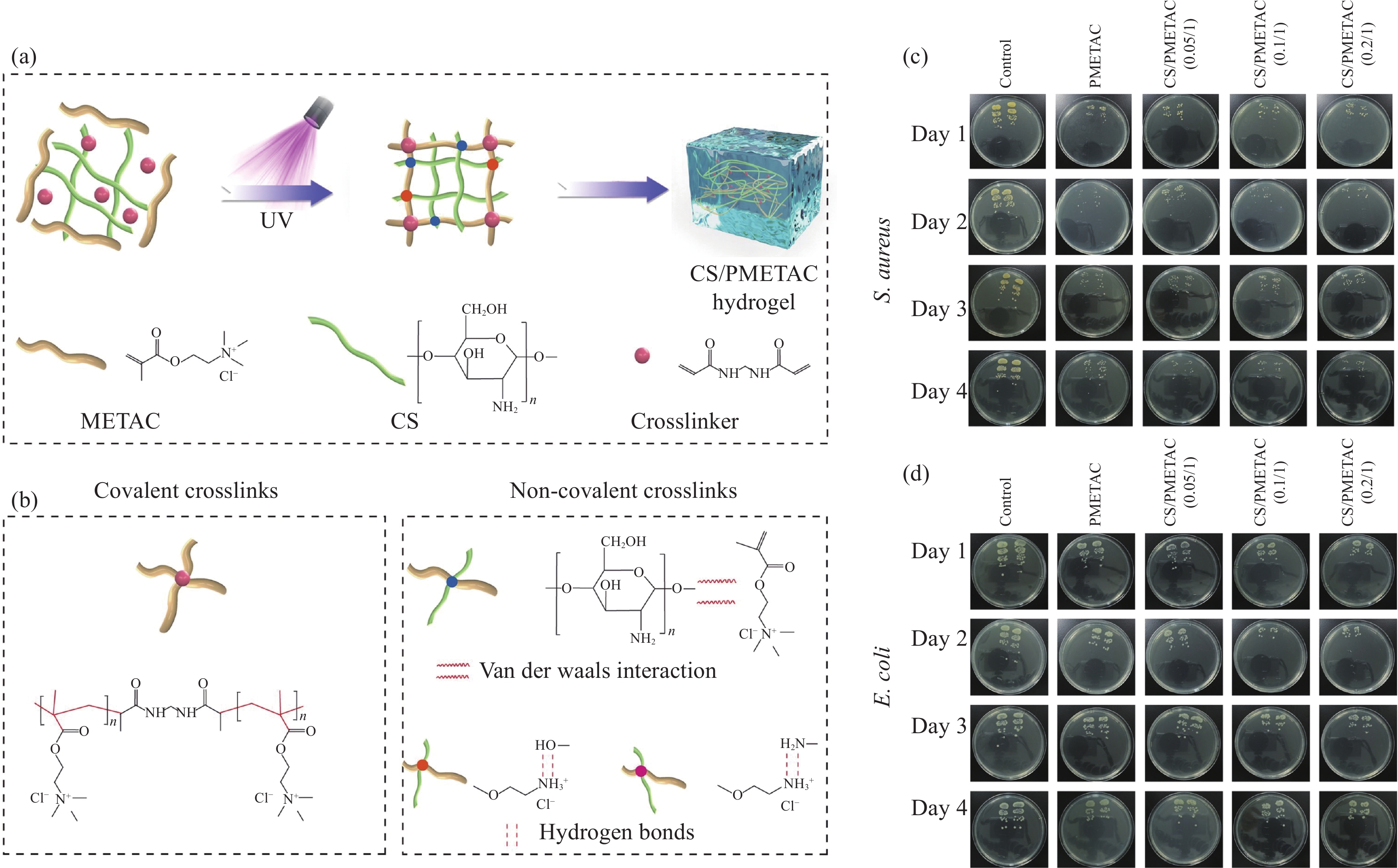
图 6 (a) 前驱体溶液经紫外照射形成CS/聚甲基丙烯酰氧基乙基三甲基氯化铵(PMETAC)水凝胶示意图[40];(b) 水凝胶交联示意图[40];((c), (d)) 对照组与CS/PMETAC水凝胶不同菌种长期抗菌图片[40]
Figure 6. (a) Schematic diagram of CS/polymethylacryloxyethyl trimethyl ammonium chloride (PMETAC) hydrogel formed by UV irradiation of precursor solutionm[40]; (b) Schematic diagram of hydrogel cross-linking[40]; ((c), (d)) Long-term antibacterial pictures of different bacteria in control group and CS/PMETAC hydrogel[40]

图 7 (a) 羧甲基壳聚糖(CMCS)/低聚原花青苷(OPC)水凝胶形成及相互作用示意图[41];(b) 水凝胶自愈机制示意图[41];(c) 水凝胶对不同菌种杀菌率及抑菌圈照片及统计图[41];(d) 空白组和水凝胶处理伤口、愈合率和定量分析肉芽组织厚度图片[42]
Figure 7. (a) Carboxymethyl chitosan (CMCS)/oligoproanthocyanidin (OPC) hydrogel formation and interaction diagram[41]; (b) Schematic diagram of the self-healing mechanism of hydrogel[41]; (c) Photos and statistical charts of bactericidal rate and bacteriostatic zone of hydrogel against different strains[41]; (d) Images of wounds, healing rates and quantitative granulation tissue thickness in blank and hydrogel dressing treatments[41]
CCS—Catechol modified chitosan ; CCHOs—aldehyde-modified cellulose nanocrystals; x in CMCS/OPCx—Content of OPC is x%

图 8 (a) 水凝胶膜形成机制及抗菌作用示意图[46];((b), (c)) 贮藏10天期间使用聚乙烯(PE)膜、再生纤维素(RC)膜和试验组(N-3)包装猪肉中细菌总数和总挥发性氮(TVB-N)[46];(d) 膜对两种菌落覆盖的琼脂图片[46];(e) 25℃不同膜对奶酪包装影响图片[46]
Figure 8. (a) Schematic diagram of hydrogel film formation mechanism and antibacterial action[46]; ((b), (c)) Total bacterial count and total volatile nitrogen (TVB-N) in pork packaged with polyethylene (PE) film, regenerated cellulose (RC) film and experimental group (N-3) during 10 days of storage[46]; (d) AGAR images of the membrane covering both colonies[46]; (e) Pictures of the effects of different films on cheese packaging at 25℃[46]
CNC—Cellulose nanocrystal; r-CNC—Rosin-grafted CNCs; PVC—Polyvinyl chloride

图 9 (a) 多功能电子皮肤制备示意图[53];(b) 菌落溶液培养24 h后图片[53];(c) 腕部脉搏随运动实时I-t曲线[53];(d) 弯曲手指关节的实时I-t曲线[53];(e) 行走时电子皮肤的实时I-t曲线[53]
Figure 9. (a) Schematic diagram of multifunctional e-skin preparation[53]; (b) Pictures after 24 h of colony solution culture[53]; (c) Real-time I-t curves of wrist pulse with movement[53]; (d) Real-time I-t curve of bending finger joint[53]; (e) Real-time I-t curve of the electronic skin during walking[53]
TOCN—TEMPO oxidized cellulose nanofibers; t—Incubation time
表 1 不同生物基抗菌水凝胶作用机制、基底材料对比分析
Table 1. Comparative analysis of action mechanism and substrate materials of different biological - based antibacterial hydrogels
Action Substrate Antimicrobial agent Antimicrobial ability Citation Other properties References Metal coordination CS Ag+ 5 mm inhibition zone Wound dressing Tensile strength (0.17 MPa) [17] Schiff base DCS, PEGSH — Lethal rate for E. coli and
S. aureus exceeds 95%Medical adhesive Blood absorption performance
((1300±50)%)[19] Metal coordination CMC, PVA AgNPs 15 mm inhibition zone against UTI pathogens Antibacterial material — [23] Silicon-oxygen
covalent bondBC SPG Inhibitory against E. coli and
S. aureusAntibacterial film — [26] Metal coordination CMS, PVA AgNPs 6 mm inhibition zone Wound dressing Swelling index 243% [29] Schiff base ASA, COS ZnO NPs 3.1 cm inhibition zone against
B. subtilisWound dressing Water vapor permeability
682 g/m2/24 h[32] Hydrogen bond ASA PL Lethal rate for E. coli and S. aureus exceeds 91.01% and 84.97% Wound healing materials PL broad-sectrum
and efficient[33] Mannich reaction BSA THPS 15 mm inhibition zone Wound healing dressing Wide alicability [37] Electrostatic
InteractionGel, CS, NF CA Lethal rate for E. coli and S. aureus exceeds 90% Wound dressing Tensile strength (0.85±0.02) MPa [38] Notes: PL—Polylysine; NF—Non-woven fabrics; SPG—Schizophyllan.  下载: 导出CSV
-
[1] CAO Z M, LUO Y E, LI Z Y, et al. Antibacterial hybrid hydrogels[J]. Macromolecular Bioscience,2021,21(1):2000252. doi: 10.1002/mabi.202000252 [2] LI S Q, DONG S J, XU W G, et al. Antibacterial hydrogels[J]. Advanced Science,2018,5(5):1700527. doi: 10.1002/advs.201700527 [3] AHMED E M. Hydrogel: Preparation, characterization, and applications: a review[J]. Journal of Advanced Research,2015,6(2):105-121. doi: 10.1016/j.jare.2013.07.006 [4] CARREÑO G, PEREIRA A, ÁVILA-SALAS F, et al. Development of “on-demand” thermo-responsive hydrogels for anti-cancer drugs sustained release: Rational design, in silico prediction and in vitro validation in colon cancer models[J]. Materials Science and Engineering: C,2021,131:112483. doi: 10.1016/j.msec.2021.112483 [5] CHENG L A, CAI Z W, YE T J, et al. Injectable polypeptide-protein hydrogels for promoting infected wound healing[J]. Advanced Functional Materials,2020,30(25):2001196. doi: 10.1002/adfm.202001196 [6] HOQUE J, BHATTACHARJEE B, PRAKASH R G, et al. Dual function injectable hydrogel for controlled release of antibiotic and local antibacterial therapy[J]. Biomacromolecules,2018,19(2):267-278. doi: 10.1021/acs.biomac.7b00979 [7] YANG K R, HAN Q, CHEN B P, et al. Antimicrobial hydrogels: Promising materials for medical application[J]. International Journal of Nanomedicine,2018,13:2217-2263. doi: 10.2147/IJN.S154748 [8] 许雨芩, 杨建军, 吴庆云, 等. 抗菌型高分子水凝胶研究进展[J]. 化工新型材料, 2022, 50(9):218-224, 228. doi: 10.19817/j.cnki.issn1006-3536.2022.09.043XU Yuqin, YANG Jianjun, WU Qingyun, et al. Research progress of antibacterial polymer hydrogels[J]. New Chemical Materials,2022,50(9):218-224, 228(in Chinese). doi: 10.19817/j.cnki.issn1006-3536.2022.09.043 [9] DUQUETTE D, DUMONT M J. Comparative studies of chemical crosslinking reactions and applications of bio-based hydrogels[J]. Polymer Bulletin,2019,76(5):2683-2710. doi: 10.1007/s00289-018-2516-6 [10] WANG W D, NARAIN R, ZENG H B. Rational design of self-healing tough hydrogels: A mini review[J]. Frontiers in Chemistry,2018,6:497. doi: 10.3389/fchem.2018.00497 [11] XIE M M, GAO M, YUN Y, et al. Antibacterial nanomaterials: Mechanisms, impacts on antimicrobial resistance and design principles[J]. Angewandte Chemie, 2023, 135(17) : e202217345. [12] SLAVIN Y N, ASNIS J, H FELI U O, et al. Metal nanoparticles: Understanding the mechanisms behind antibacterial activity[J]. Journal of Nanobiotechnology,2017,15(1): 65:1. doi: 10.1186/s12951-016-0241-6 [13] 管瑛, 恽亮, 韦恩泽, 等. 抗菌材料的研究现状[J]. 生物化工, 2022, 8(2):164-166. doi: 10.3969/j.issn.2096-0387.2022.02.043GUAN Ying, YUN Liang, WEI Enze, et al. Research status of antibacterial materials[J]. Biological Chemical Engineering,2022,8(2):164-166(in Chinese). doi: 10.3969/j.issn.2096-0387.2022.02.043 [14] MURUGESAN S, SCHEIBEL T. Chitosan-based nanocomposites for medical applications[J]. Journal of Polymer Science,2021,59(15):1610-1642. doi: 10.1002/pol.20210251 [15] MOEINI A, PEDRAM P, MAKVANDI P, et al. Wound healing and antimicrobial effect of active secondary metabolites in chitosan-based wound dressings: A review[J]. Carbohydrate Polymers,2020,233:115839. doi: 10.1016/j.carbpol.2020.115839 [16] REZAEI N, HAMIDABADI H G, KHOSRAVIMELAL S, et al. Antimicrobial peptides-loaded smart chitosan hydrogel: Release behavior and antibacterial potential against antibiotic resistant clinical isolates[J]. International Journal of Biological Macromolecules,2020,164:855-862. doi: 10.1016/j.ijbiomac.2020.07.011 [17] LI P, ZHAO J, CHEN Y, et al. Preparation and characterization of chitosan physical hydrogels with enhanced mechanical and antibacterial properties[J]. Carbohydrate Polymers,2017,157:1383-1392. doi: 10.1016/j.carbpol.2016.11.016 [18] WANG J L, ZHUANG S T. Chitosan-based materials: preparation, modification and application[J]. Journal of Cleaner Production, 2022, 355: 131825. [19] SONG F Y, KONG Y, SHAO C Y, et al. Chitosan-based multifunctional flexible hemostatic bio-hydrogel[J]. Acta Biomaterialia,2021,136:170-183. doi: 10.1016/j.actbio.2021.09.056 [20] CHEN Z, YAO J P, ZHAO J L, et al. Injectable wound dressing based on carboxymethyl chitosan triple-network hydrogel for effective wound antibacterial and hemostasis[J]. International Journal of Biological Macromolecules,2023,225:1235-1245. doi: 10.1016/j.ijbiomac.2022.11.184 [21] ZAINAL S H, MOHD N H, SUHAILI N, et al. Preparation of cellulose-based hydrogel: A review[J]. Journal of Materials Research and Technology,2021,10:935-952. doi: 10.1016/j.jmrt.2020.12.012 [22] GOMRI C, CRETIN M, SEMSARILAR M. Recent progress on chemical modification of cellulose nanocrystal (CNC) and its application in nanocomposite films and membranes-A comprehensive review[J]. Carbohydrate Polymers, 2022, 294:119790. [23] ALSHEHRI S M, ALDALBAHI A, AL-HAJJI A B, et al. Development of carboxymethyl cellulose-based hydrogel and nanosilver composite as antimicrobial agents for UTI pathogens[J]. Carbohydrate Polymers,2016,138:229-236. doi: 10.1016/j.carbpol.2015.11.004 [24] PENG N, WANG Y F, YE Q F, et al. Biocompatible cellulose-based superabsorbent hydrogels with antimicrobial activity[J]. Carbohydrate Polymers,2016,137:59-64. doi: 10.1016/j.carbpol.2015.10.057 [25] 孙振炳, 李晓宝, 姚曜, 等. 细菌纤维素抗菌复合材料的制备和应用[J]. 包装工程, 2021, 42(13):21-28.SUN Zhenbing, LI Xiaobao, YAO Yao, et al. Preparation and application of bacterial cellulose antibacterial composite material[J]. Packaging Engineering,2021,42(13):21-28(in Chinese). [26] HAMEDI S, SHOJAOSADATI S A, NAJAFI V, et al. A novel double-network antibacterial hydrogel based on aminated bacterial cellulose and schizophyllan[J]. Carbohydrate Polymers,2020,229:115383. doi: 10.1016/j.carbpol.2019.115383 [27] CUI C L, JIA Y Z, SUN Q, et al. Recent advances in the preparation, characterization, and food application of starch-based hydrogels[J]. Carbohydrate Polymers, 2022, 291: 119624. [28] VILLANUEVA M E, DIEZ A M, GOMZALEZ J A, et al. Antimicrobial activity of starch hydrogel incorporated with copper nanoparticles[J]. ACS Applied Materials & Interfaces, 2016, 8(25): 16280-16288. [29] OUNKAEW A, KASEMSIRI P, JETSRISUPARB K, et al. Synthesis of nanocomposite hydrogel based carboxymethyl starch/polyvinyl alcohol/nanosilver for biomedical materials[J]. Carbohydrate Polymers,2020,248:116767. doi: 10.1016/j.carbpol.2020.116767 [30] YUAN L, WU Y, GU Q S, et al. Injectable photo crosslinked enhanced double-network hydrogels from modified sodium alginate and gelatin[J]. International Journal of Biological Macromolecules,2017,96:569-577. doi: 10.1016/j.ijbiomac.2016.12.058 [31] ARAFA E G, SABAA M W, MOHAMED R R, et al. Eco-friendly and biodegradable sodium alginate/quaternized chitosan hydrogel for controlled release of urea and its antimicrobial activity[J]. Carbohydrate Polymers,2022,291:119555. doi: 10.1016/j.carbpol.2022.119555 [32] ZHANG M, QIAO X N, HAN W W, et al. Alginate-chitosan oligosaccharide-ZnO composite hydrogel for accelerating wound healing[J]. Carbohydrate Polymers,2021,266:118100. doi: 10.1016/j.carbpol.2021.118100 [33] JIN F Y, LIAO S Q, LI W, et al. Amphiphilic sodium alginate-polylysine hydrogel with high antibacterial efficiency in a wide pH range[J]. Carbohydrate Polymers,2023,299:120195. doi: 10.1016/j.carbpol.2022.120195 [34] BUHRMAN J S, COOK L C, RAYAHIN J E, et al. Proteolytically activated anti-bacterial hydrogel microspheres[J]. Journal of Controlled Release,2013,171(3):288-295. doi: 10.1016/j.jconrel.2013.06.023 [35] ZHOU J J, ZHANG H R, FAREED M S, et al. An injectable peptide hydrogel constructed of natural antimicrobial peptide J-1 and ADP shows anti-infection, hemostasis, and antiadhesion efficacy[J]. ACS Nano,2022,16(5):7636-7650. doi: 10.1021/acsnano.1c11206 [36] MAYOL G F, DEFELIPE L A, ARCON J P, et al. Solvent sites improve docking performance of protein–protein complexes and protein–protein interface-targeted drugs[J]. Journal of Chemical Information and Modeling,2022,62(15):3577-3588. doi: 10.1021/acs.jcim.2c00264 [37] OUYANG J, BU Q Y, TAO N, et al. A facile and general method for synthesis of antibiotic-free protein-based hydrogel: Wound dressing for the eradication of drug-resistant bacteria and biofilms[J]. Bioactive Materials,2022,18:446-458. doi: 10.1016/j.bioactmat.2022.03.033 [38] WANG L L, LI D W, SHEN Y, et al. Preparation of Centella asiatica loaded gelatin/chitosan/nonwoven fabric composite hydrogel wound dressing with antibacterial property[J]. International Journal of Biological Macromolecules,2021,192:350-359. doi: 10.1016/j.ijbiomac.2021.09.145 [39] JIA B, LI G W, CAO E T, et al. Recent progress of antibacterial hydrogels in wound dressings[J]. Materials Today Bio, 2023, 19: 100582. [40] YU Q, YAN Y G, HUANG J, et al. A multifunctional chitosan-based hydrogel with self-healing, antibacterial, and immunomodulatory effects as wound dressing[J]. International Journal of Biological Macromolecules, 2023, 231: 123149. [41] HE Y M, GUO S, CHANG R, et al. Facile preparation of antibacterial hydrogel with multi-functions based on carboxymethyl chitosan and oligomeric procyanidin[J]. RSC Advances,2022,12(32):20897-20905. doi: 10.1039/D2RA04049B [42] LU H B, LI X L, ZHANG M, et al. Antibacterial cellulose nanocrystal-Incorporated hydrogels with satisfactory vascularization for enhancing skin regeneration[J]. Frontiers in Bioengineering and Biotechnology, 2022, 10: 876936. [43] YOU S Y, XIANG Y J, QI X L, et al. Harnessing a biopolymer hydrogel reinforced by copper/tannic acid nanosheets for treating bacteria-infected diabetic wounds[J]. Materials Today Advances,2022,15:100271. doi: 10.1016/j.mtadv.2022.100271 [44] PRIYADARSHI R, ROY S, GHOSH T, et al. Antimicrobial nanofillers reinforced biopolymer composite films for active food packaging applications-a review[J]. Sustainable Materials and Technologies,2022,32:e00353. doi: 10.1016/j.susmat.2021.e00353 [45] PURWAR R, VERMA A, BATRA R. Antimicrobial gelatin/sericin/clay films for packaging of hygiene products[J]. Journal of Polymer Engineering,2019,39(8):744-751. doi: 10.1515/polyeng-2018-0406 [46] WU Y H, LI Q, ZHANG X Z, et al. Cellulose-based peptidopolysaccharides as cationic antimicrobial package films[J]. International Journal of Biological Macromolecules,2019,128:673-680. doi: 10.1016/j.ijbiomac.2019.01.172 [47] DHIVYA A, AMIT K. Pineapple juice clarification by continuous dead-end microfiltration using a low-cost ceramic membrane[J]. Journal of Food Measurement and Characterization,2023,17(1):863-881. doi: 10.1007/s11694-022-01634-5 [48] SONG Z, MA T, ZHI X, et al. Cellulosic films reinforced by chitosan-citric complex for meat preservation: Influence of nonenzymatic browning[J]. Carbohydrate Polymers,2021,272:118476. doi: 10.1016/j.carbpol.2021.118476 [49] LEITE L S F, BILATTO S, PASCHOALIN R T, et al. Eco-friendly gelatin films with rosin-grafted cellulose nanocrystals for antimicrobial packaging[J]. International Journal of Biological Macromolecules,2020,165:2974-2983. doi: 10.1016/j.ijbiomac.2020.10.189 [50] ZHAO H, LIU M, ZHANG Y, et al. Nanocomposite hydrogels for tissue engineering applications[J]. Nanoscale,2020,12(28):14976-14995. doi: 10.1039/D0NR03785K [51] LIN F C, WANG Z, SHEN Y P, et al. Natural skin-inspired versatile cellulose biomimetic hydrogels[J]. Journal of Materials Chemistry A,2019,7(46):26442-26455. doi: 10.1039/C9TA10502F [52] SHI Y P, WEI X L, WANG K M, et al. Integrated all-fiber electronic skin toward self-powered sensing sports systems[J]. ACS Applied Materials & Interfaces,2021,13(42):50329-50337. [53] XU H, LIU D, SONG Y, et al. Ultra-sensitive and flexible electronic skin from nanocellulose/AgNWs hydrogel films with highly transparent, antibacterial and electromagnetic shielding properties[J]. Composites Science and Technology,2022,228:109679. doi: 10.1016/j.compscitech.2022.109679 -

 下载:
下载:


 点击查看大图

计量
- 文章访问数: 777
- HTML全文浏览量: 403
- PDF下载量: 70
- 被引次数: 0
