


Research progress on hydroxyapatite/graphene oxide composite scaffolds in the treatment of bone defect repair
-
摘要: 骨缺损引起的骨疾病是现代医学非常棘手的一种疾病,正常的骨组织发生病变或外部因素的物理损伤都会形成难愈合的骨缺损;骨缺损部位复杂的微环境决定其治疗条件也更苛刻。构建可以用于骨缺损的支架是骨组织工程技术的一个重要方向,也是解决骨缺损疾病难题的重要手段。本文简单介绍了羟基磷灰石/氧化石墨烯(HA/GO)复合材料的制备原理和方法,细胞实验表明HA/GO对骨细胞没有明显的毒性,粗糙的表面结构更有利于骨细胞的增殖分化,在此基础上详细介绍了用于治疗骨缺损疾病HA/GO复合支架的构建方式及其性能。最后讨论了HA/GO复合支架在骨疾病治疗中的发展前景和面临的挑战。Abstract: Bone defects caused by bone diseases are a very challenging issue in modern medicine. Both pathological changes in normal bone tissue and physical damage caused by external factors can lead to difficult-to-heal bone defects. The complex microenvironment of the bone defect site makes treatment conditions more demanding. Constructing scaffolds for bone defects is an important direction in bone tissue engineering technology and a crucial means to solve the problem of bone defect diseases. This article briefly introduces the preparation principles and methods of hydroxyapatite/graphene oxide (HA/GO) composite materials. HA/GO has no apparent toxicity to bone cells, and its rough surface structure is more conducive to the proliferation and differentiation of bone cells. Based on this, the construction methods and performance of HA/GO composite scaffolds for treating bone defect diseases are described in detail. Finally, the development prospects and challenges of HA/GO composite scaffolds in the treatment of bone diseases are discussed.
-
Key words:
- graphene oxide /
- hydroxyapatite /
- bone diseases /
- composite brackets /
- tissue engineering
-


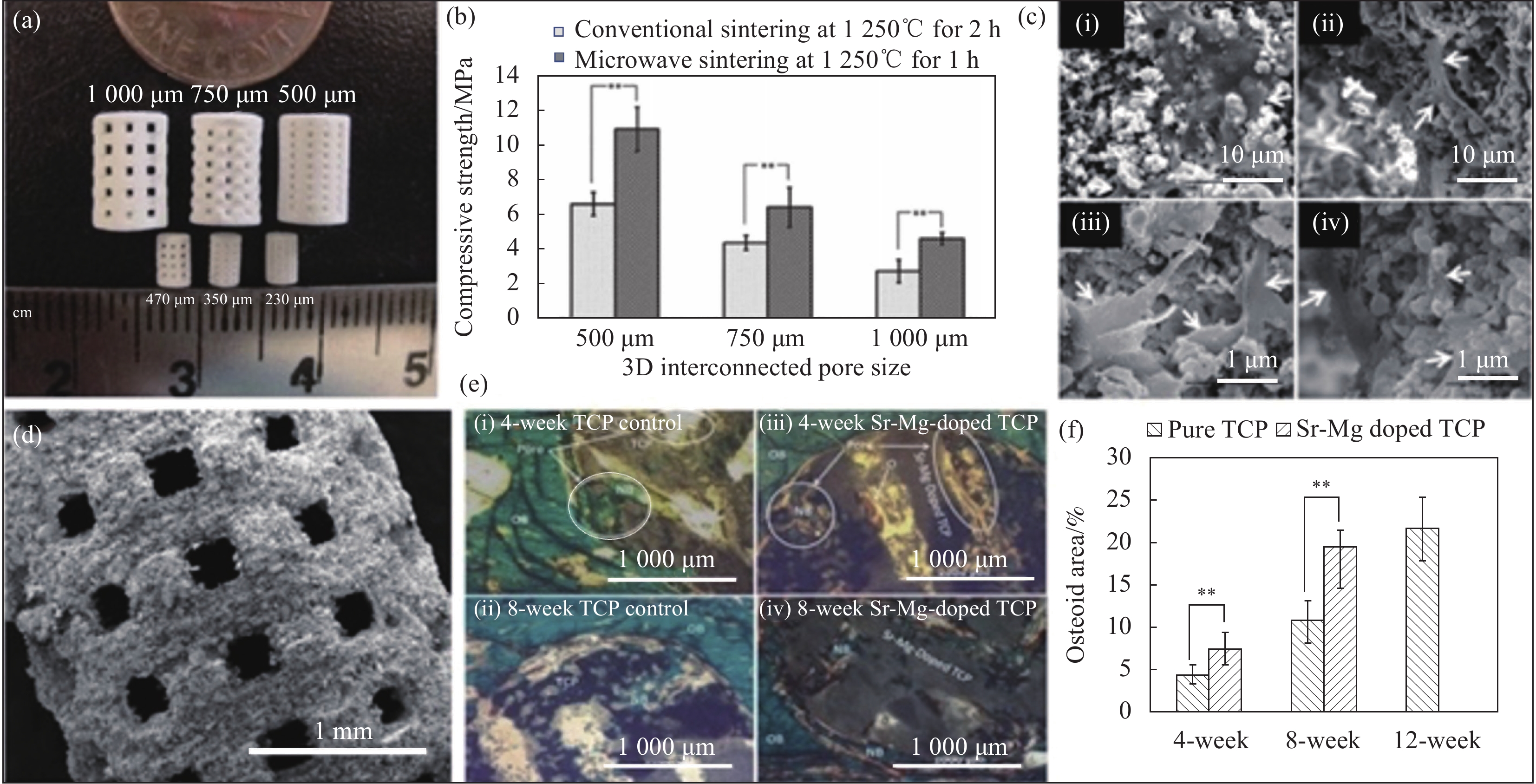
图 4 具有不同孔径大小的3D打印支架[30-32]:(a) 3D打印磷酸三钙(TCP)支架;(b) 1250℃烧结支架的抗压强度比较;(c) 细胞在支架表面和3D互连大孔内的黏附和增殖;(d) 纯TCP支架的SEM图像;(e) 植入物的显微照片;(f) 类骨面积分数的组织形态计量学分析
Figure 4. 3D printed brackets with different aperture sizes[30-32]: (a) 3D printing tricalcium phosphate (TCP) scaffolds; (b) Comparison of compressive strength of 1250℃ sintered scaffolds; (c) Adhesion and proliferation of cells on the surface of scaffolds and within 3D interconnected macropores; (d) SEM image of pure TCP scaffolds; (e) Microscopic photos of implants; (f) Histomorphometric analysis of osteoid area fraction

图 2 不同构建方式HA/GO复合支架形貌:(a) 壳聚糖基复合支架[21];(b) 明胶基复合支架[22];(c) 纳米纤维基复合支架[29];(d) 自组装法制备的HA/GO复合支架[35]
Figure 2. HA/GO composite scaffold morphology with different construction methods: (a) Chitosan based composite scaffold[21]; (b) Gelatin based composite scaffold[22]; (c) Nanofiber based composite scaffold[29]; (d) HA/GO composite scaffold prepared by self-assembly method[35]
GHA—Gelatin-HA scaffold; GOGHA0.5—GHA scaffold with 0.5wt%GO; nHA—Nano-hydroxyapatite; rGO—Reduced graphene oxide


图 6 HA/GO复合支架的力学性能:(a) 不同聚己内酯(PCL)含量的HA/GO应力-应变行为[5];(b) 不同含量HA/GO纳米压痕测试[18];(c) 不同海藻酸钠(SA)含量的应力-应变曲线[39]
Figure 6. Mechanical properties of HA/GO composite scaffolds: (a) Stress-strain behavior of HA/GO with different poly(ε-caprolactone) (PCL) contents[5]; (b) HA/GO nanoindentation testing with different contents[18]; (c) Stress-strain curves of different sodium alginate (SA) contents[39]
HAP—Hydroxyapatite; Gs—Graphene nanosheets; SHA—Spherical porous hydroxyapatite; M—Elastic modulus; H—Hardness; σe1*—Elastic collapse stress; E*—Calculate the linear elastic modulus; Δσ/Δε—Collapse plateau modulus; εe1*—Elastic collapse strain

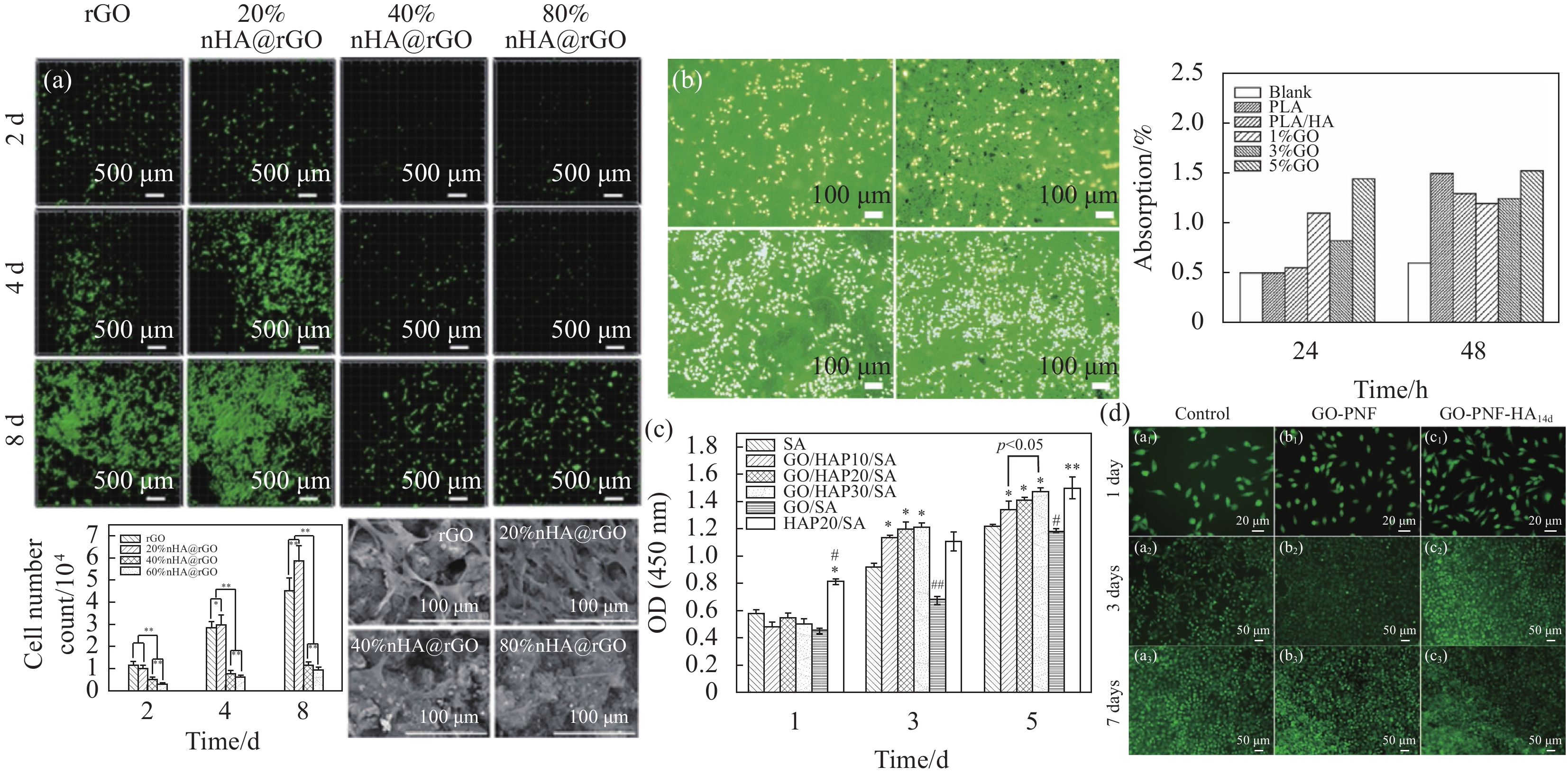
图 7 HA/GO复合支架的细胞毒性[2, 4, 18, 33, 40]:(a) 小鼠胚胎成骨细胞前体细胞 (MC3T3-E1)在不同材料上的噻唑蓝检测 (MTT)检测结果;(b) Saos-2成骨样细胞在不同材料上的细胞增殖率;(c) 人骨髓间充斥干细胞 (hMSCs)在不同材料上的细胞活性;(d) 人骨肉瘤细胞(MG-63)在不同材料上的增殖情况;(e) hMSCs细胞代谢情况;(f) 不同HA含量的复合材料形貌;(g) MC3T3-E1细胞形态;(h) MC3T3-E1细胞的增殖情况;(i) 培养不同天数MC3T3-E1细胞细胞增值状况
Figure 7. Cytotoxicity of HA/GO composite scaffolds[2, 4, 18, 33, 40]: (a) 3-[4, 5-dimethylthiazol-2-yl]-2, 5-diphenyltetrazolium bromide (MTT) monitoring results of mouse osteoblast (MC3T3-E1) cells on different materials; (b) Cell proliferation rate of Saos-2 osteoblast like cells on different materials; (c) Cellular activity of human mesenchymal stem cells (hMSCs) cells on different materials; (d) The proliferation of Human osteosarcoma cells (MG-63) cells on different materials; (e) Metabolism of hMSCs cells; (f) Morphology of composite materials with different HA contents; (g) MC3T3-E1 cell morphology; (h) The proliferation of MC3T3-E1 cells; (i) Cell proliferation status of MC3T3-E1 cells cultured for different days
OD—Optical density; CTR—Control group; SBF—Simulated body fluid; FGO—Functional GO; FGHA—Functional GO/HA; RHA—Rod HA; SHA—Spherical HA

图 8 HA/GO复合支架的细胞增殖附着实验:(a)大鼠骨髓间充质干细胞(rBMSCs)活细胞密度[33];(b) MC3T3-E1细胞增值情况[29];(c) 人骨肉瘤细胞(MG-63)细胞增值情况[39];(d) MC3T3-E1细胞增值情况[50]
Figure 8. Cell proliferation and attachment assay for HA/GO composite scaffolds: (a) Rat bone mesenchymal stem cells (rBMSCs) live cell density[33]; (b) MC3T3-E1 cell proliferation status[29]; (c) Human osteosarcoma cells (MG-63) proliferation status[39]; (d) MC3T3-E1 cell proliferation status[50]
p—Significance level; PLA—Polylactic acid

-
[1] 杨洪, 张昊烨, 陈新艳. 氧化石墨烯增强磷酸钙生物水泥[J]. 复合材料学报, 2016, 33(4):852-858. doi: 10.13801/j.cnki.fhclxb.20150915.001YANG Hong, ZHANG Haoye, CHEN Xinyan. Calcium phosphate biocements reinforced with graphene oxide[J]. Acta Materiae Compositae Sinica,2016,33(4):852-858(in Chinese). doi: 10.13801/j.cnki.fhclxb.20150915.001 [2] RAMANI D, SASTRY T P. Bacterial cellulose-reinforced hydroxyapatite functionalized graphene oxide: A potential osteoinductive composite[J]. Cellulose,2014,21(5):3585-3595. doi: 10.1007/s10570-014-0313-4 [3] FIROOZABADI F D, SAADATABADI A R, ASEFNEJAD A. Fabrication and evaluation of in vitro studies of biodegradable and antibacterial composite scaffolds based on polylactic acid-polycaprolactone-hdroxyapatite reinforced with graphene and zinc oxide nanoparticles for use in orthopedic surgery[J]. Iranian Journal of Materials Science and Engineering,2022,19(2):1-19. [4] RAUCCI M G, GIUGLIANO D, LONGO A, et al. Comparative facile methods for preparing graphene oxide-hydroxyapatite for bone tissue engineering[J]. Journal of Tissue Engineering and Regenerative Medicine,2017,11(8):2204-2216. doi: 10.1002/term.2119 [5] ZHOU K Q, GAO R, JIANG S H. Morphology, thermal and mechanical properties of poly (epsilon-caprolactone) biocomposites reinforced with nano-hydroxyapatite decorated graphene[J]. Journal of Colloid and Interface Science,2017,496:334-342. doi: 10.1016/j.jcis.2017.02.038 [6] RAMADAS M, BHARATH G, PONPANDIAN N, et al. Investigation on biophysical properties of hydroxyapatite/graphene oxide (HAp/GO) based binary nanocomposite for biomedical applications[J]. Materials Chemistry and Physics,2017,199:179-184. doi: 10.1016/j.matchemphys.2017.07.001 [7] YAO C, ZHU J, XIE A, et al. Graphene oxide and creatine phosphate disodium dual template-directed synthesis of GO/hydroxyapatite and its application in drug delivery[J]. Materials Science & Engineering C,2017,73:709-715. doi: 10.1016/j.msec.2016.11.083 [8] RODRIGUEZ-GONZALEZ C, SALAS P, LOPEZ-MARIN L M, et al. Hydrothermal synthesis of graphene oxide/multiform hydroxyapatite nanocomposite: Its influence on cell cytotoxicity[J]. Materials Research Express,2018,5(12):125023. doi: 10.1088/2053-1591/aae29c [9] ZENG Y, PEI X, YANG S, et al. Graphene oxide/hydroxyapatite composite coatings fabricated by electrochemical deposition[J]. Surface & Coatings Technology,2016,286:72-79. [10] LI M, WANG Y, LIU Q, et al. In situ synthesis and biocompatibility of nano hydroxyapatite on pristine and chitosan functionalized graphene oxide[J]. Journal of Materials Chemistry B,2013,1(4):475-484. doi: 10.1039/C2TB00053A [11] LIU H, XI P, XIE G, et al. Simultaneous reduction and surface functionalization of graphene oxide for hydroxyapatite mineralization[J]. Journal of Physical Chemistry C,2012,116(5):3334-3341. doi: 10.1021/jp2102226 [12] GIROLAMI G S, JENSEN J A, POLLINA D M, et al. Organometallic route to the chemical vapor deposition of titanium carbide films at exceptionally low temperatures[J]. Journal of the American Chemical Society,1987,109(5):1579-1580. doi: 10.1021/ja00239a053 [13] KUDIN K N, OZBAS B, SCHNIEPP H C, et al. Raman spectra of graphite oxide and functionalized graphene sheets[J]. Nano Letters,2008,8(1):36-41. doi: 10.1021/nl071822y [14] CHENG M, YANG R, ZHANG L, et al. Restoration of graphene from graphene oxide by defect repair[J]. Carbon,2012,50(7):2581-2587. doi: 10.1016/j.carbon.2012.02.016 [15] LIU Y, DANG Z, WANG Y, et al. Hydroxyapatite/graphene-nanosheet composite coatings deposited by vacuum cold spraying for biomedical applications: Inherited nanostructures and enhanced properties[J]. Carbon,2014,67:250-259. doi: 10.1016/j.carbon.2013.09.088 [16] DUAN P, SHEN J, ZOU G, et al. Biomimetic mineralization and cytocompatibility of nanorod hydroxyapatite/graphene oxide composites[J]. Frontiers of Chemical Science and Engineering,2018,12(4):798-805. doi: 10.1007/s11705-018-1708-9 [17] RADHA G, VINOD R K, VENKATESAN B, et al. In vitro studies of graphene oxide reinforced hydroxyapatite nanobiocomposite on human erythrocytes[C]. Dae Solid State Physics Symposium 2016. Melville: AIP Publishing LLC, 2017. [18] DUAN P, SHEN J, ZOU G, et al. Synthesis spherical porous hydroxyapatite/graphene oxide composites by ultrasonic-assisted method for biomedical applications[J]. Biomedical Materials,2018,13(4):045001. doi: 10.1088/1748-605X/aab3ea [19] 熊伟, 袁灵梅, 钱国文, 等. 临界骨缺损动物模型评估骨组织工程支架成骨效能的价值[J]. 中国组织工程研究, 2023, 27(35): 5714-5720.XIONG Wei, YUAN Lingmei, QIAN Guowen, et al. Value of a critical bone defect animal model in evaluating osteogenic efficacy of bone tissue engineering scaffold[J]. Chinese Journal of Tissue Engineering Research, 2023, 27(35): 5714-5720(in Chinese) [20] BHARATH G, MADHU R, CHEN S M, et al. Enzymatic electrochemical glucose biosensors by mesoporous 1D hydroxyapatite-on-2D reduced graphene oxide[J]. Journal of Materials Chemistry B,2015,3(7):1360-1370. doi: 10.1039/C4TB01651C [21] HARLEY B A, LEUNG J H, SILVA E C C M, et al. Mechanical characterization of collagen-glycosaminoglycan scaffolds[J]. Acta Biomaterialia,2007,3(4):463-474. doi: 10.1016/j.actbio.2006.12.009 [22] NAIR M, NACY D, KRISHNAN A G, et al. Graphene oxide nnoflakes incorporated gelatin-hydroxyapatite scaffolds enhance osteogenic differentiation of human mesenchymal stem cells[J]. Nanotechnology,2015,26(16):161001. doi: 10.1088/0957-4484/26/16/161001 [23] SUN B, LONG Y Z, ZHANG H D, et al. Advances in three-dimensional nanofibrous macrostructures via electrospinning[J]. Progress in Polymer Science,2014,39(5):862-890. doi: 10.1016/j.progpolymsci.2013.06.002 [24] REPANAS A, ANDRIOPOULOU S, GLASMACHER B. The significance of electrospinning as a method to create fibrous scaffolds for biomedical engineering and drug delivery applications[J]. Journal of Drug Delivery Science and Technology,2016,31:137-146. doi: 10.1016/j.jddst.2015.12.007 [25] SILL T J, VON RECUM H A. Electrospinning: Applications in drug delivery and tissue engineering[J]. Biomaterials,2008,29(13):1989-2006. doi: 10.1016/j.biomaterials.2008.01.011 [26] TAYLOR G. Electrically driven jets[J]. Proceedings of the Royal Society of London, Series A (Mathematical and Physical Sciences),1969,313(1515):453-475. [27] OKUTAN N, TERZI P, ALTAY F. Affecting parameters on electrospinning process and characterization of electrospun gelatin nanofibers[J]. Food Hydrocolloids,2014,39:19-26. doi: 10.1016/j.foodhyd.2013.12.022 [28] LIANG H P, WANG Y B, SU Z, et al. Electrospinning gelatin/chitosan/hydroxyapatite/graphene oxide composite nanofibers with antibacterial properties[J]. Journal of Inorganic Materials,2015,30(5):516-522. doi: 10.15541/jim20140528 [29] MA H, SU W, TAI Z, et al. Preparation and cytocompatibility of polylactic acid/hydroxyapatite/graphene oxide nanocomposite fibrous membrane[J]. Chinese Science Bulletin,2012,57(23):3051-3058. doi: 10.1007/s11434-012-5336-3 [30] BOSE S, VAHABZADEH S, BANDYOPADHYAY A. Bone tissue engineering using 3D printing[J]. Materials Today,2013,16(12):496-504. doi: 10.1016/j.mattod.2013.11.017 [31] TARAFDER S, BALLA V K, DAVIES N M, et al. Microwave-sintered 3D printed tricalcium phosphate scaffolds for bone tissue engineering[J]. Journal of Tissue Engineering and Regenerative Medicine,2013,7(8):631-641. doi: 10.1002/term.555 [32] BECKER S T, BOLTE H, SCHÜNEMANN K, et al. Endocultivation: The influence of delayed vs. simultaneous application of BMP-2 onto individually formed hydroxyapatite matrices for heterotopic bone induction[J]. International Journal of Oral and Maxillofacial Surgery,2012,41(9):1153-1160. doi: 10.1016/j.ijom.2012.03.031 [33] NIE W, PENG C, ZHOU X, et al. Three-dimensional porous scaffold by self-assembly of reduced graphene oxide and nano-hydroxyapatite composites for bone tissue engineering[J]. Carbon,2017,116:325-337. doi: 10.1016/j.carbon.2017.02.013 [34] XIE X, HU K, FANG D, et al. Graphene and hydroxyapatite self-assemble into homogeneous, free standing nanocomposite hydrogels for bone tissue engineering[J]. Nanoscale,2015,7(17):7992-8002. doi: 10.1039/C5NR01107H [35] 肖鑫, 张菱素, 施冬健, 等. 近红外响应性可复写光子凝胶的构筑与性能调控[J]. 高分子学报, 2022, 53(8): 933-941.XIAO Xin, ZHANG Lingsu, SHI Dongjian, et al. Construction and property control of near-infrared responsive reproducible photonic gel[J]. Acta Polymerica Sinica, 2022, 53(8): 933-941(in Chinese). [36] 王远亮, 蔡绍皙. 生物力学与骨组织工程[J]. 力学进展, 1999(2):89-100. doi: 10.6052/1000-0992-1999-2-J1998-075WANG Yuanliang, CAI Shaoxi. Biomechanics and bone tissue engineering[J]. Advances in Mechanics,1999(2):89-100(in Chinese). doi: 10.6052/1000-0992-1999-2-J1998-075 [37] 莫湘涛. 骨组织工程支架材料的降解和生物力学特性[J]. 医用生物力学, 2004(1):56-60. doi: 10.16156/j.1004-7220.2004.01.015MO Xiangtao. Degradation and biomechanical properties of bone tissue engineering scaffolds[J]. Medical Biomechanics,2004(1):56-60(in Chinese). doi: 10.16156/j.1004-7220.2004.01.015 [38] 王身国. 组织工程细胞支架[J]. 中国康复理论与实践, 2002(5):15-17. doi: 10.3969/j.issn.1006-9771.2002.05.004WANG Shenguo. Tissue engineering cell scaffold[J]. China Rehabilitation Theory & Practice,2002(5):15-17(in Chinese). doi: 10.3969/j.issn.1006-9771.2002.05.004 [39] XIONG G, LUO H, ZUO G, et al. Novel porous graphene oxide and hydroxyapatite nanosheets-reinforced sodium alginate hybrid nanocomposites for medical applications[J]. Materials Characterization,2015,107:419-425. doi: 10.1016/j.matchar.2015.07.016 [40] 段佩珍. 羟基磷灰石/氧化石墨烯复合材料的制备及其性能研究[D]. 绵阳: 西南科技大学, 2018.DUAN Peizhen. Study on the preparation and properties ofhydroxyapatite/graphene oxide composites[D]. Mianyang: Southwest University of Science and Technology, 2018. [41] 仲蕾蕾, 杨冰, 黄晓斌, 等. OPG/RANKL/RANK系统在成骨细胞和破骨细胞相互调节中的作用[J]. 中国骨质疏松杂志, 2011, 17(11):1010-1013. doi: 10.3969/j.issn.1006-7108.2011.11.018ZHONG Leilei, YANG Bing, HUANG Xiaobin, et al. The role of OPG/RANKL/RANK system in the mutual regulation of osteoblasts and osteoclasts[J]. Chinese Journal of Osteoporosis,2011,17(11):1010-1013(in Chinese). doi: 10.3969/j.issn.1006-7108.2011.11.018 [42] 李秀森, 郭子宽, 杨靖清, 等. 骨髓间充质干细胞的生物学特性[J]. 解放军医学杂志, 2000(5):346-348. doi: 10.3321/j.issn:0577-7402.2000.05.011LI Xiusen, GUO Zikuan, YANG Jingqing, et al. Biological characteristics of bone marrow mesenchymal stem cells[J]. Chinese Journal of Medicine,2000(5):346-348(in Chinese). doi: 10.3321/j.issn:0577-7402.2000.05.011 [43] STANKOVICH S, PINER R D, NGUYEN S T, et al. Synthesis and exfoliation of isocyanate-treated graphene oxide nanoplatelets[J]. Carbon,2006,44(15):3342-3347. doi: 10.1016/j.carbon.2006.06.004 [44] SOTOME S, UEMURA T, KIKUCHI M, et al. Synthesis and in vivo evaluation of a novel hydroxyapatite/collagen-alginate as a bone filler and a drug delivery carrier of bone morphogenetic protein[J]. Materials Science and Engineering: C,2004,24(3):341-347. doi: 10.1016/j.msec.2003.12.003 [45] PETER M, GANESH N, SELVAMURUGAN N, et al. Preparation and characterization of chitosan-gelatin/nanohydroxyapatite composite scaffolds for tissue engineering applications[J]. Carbohydrate Polymers,2010,80(3):687-694. doi: 10.1016/j.carbpol.2009.11.050 [46] LIN H R, YEH Y J. Porous alginate/hydroxyapatite composite scaffolds for bone tissue engineering: Preparation, characterization, and in vitro studies[J]. Journal of Biomedical Materials Research Part B-Applied Biomaterials,2004,71B(1):52-65. doi: 10.1002/jbm.b.30065 [47] DEPAN D, GIRASE B, SHAH J S, et al. Structure-process-property relationship of the polar graphene oxide-mediated cellular response and stimulated growth of osteoblasts on hybrid chitosan network structure nanocomposite scaffolds[J]. Acta Biomaterialia,2011,7(9):3432-3445. doi: 10.1016/j.actbio.2011.05.019 [48] D'LIMA D D, LEMPERLE S M, CHEN P C, et al. Bone response to implant surface morphology[J]. The Journal of Arthroplasty,1998,13(8):928-934. doi: 10.1016/S0883-5403(98)90201-7 [49] BAKR A M, ANIS B, EL HOTABY W. Sounchemical synthesis of graphene/nano hydroxyapatite composites for potential biomedical application[J]. Egyptian Journal of Chemistry,2022,65(2):669-678. doi: 10.21608/EJCHEM.2021.91241.4339 [50] WANG J, OUYANG Z, REN Z, et al. Self-assembled peptide nanofibers on graphene oxide as a novel nanohybrid for biomimetic mineralization of hydroxyapatite[J]. Carbon,2015,89:20-30. doi: 10.1016/j.carbon.2015.03.024 [51] 陈何维, 袁波, 王林楠, 等. 羟基磷灰石/氧化石墨烯/氢氧化镁复合涂层镁-钙合金的耐蚀性及骨折修复效果研究[J]. 稀有金属材料与工程, 2021, 50(10):3698-3706.CHEN Hewei, YUAN Bo, WANG Linnan, et al. Study on the corrosion resistance and fracture repair effect of magnesium-calcium alloy coated with hydroxyapatite/GO/magnesium hydroxide composite coating[J]. Rare Metal Materials and Engineering,2021,50(10):3698-3706(in Chinese). [52] GUZMAN R, NARDECCHIA S, GUTIERREZ M C, et al. Chitosan scaffolds containing calcium phosphate salts and rhBMP-2: In vitro and in vivo testing for bone tissue regeneration[J]. Plos One,2014,9(2):e87149. doi: 10.1371/journal.pone.0087149 -

 下载:
下载:


 点击查看大图

计量
- 文章访问数: 531
- HTML全文浏览量: 255
- PDF下载量: 29
- 被引次数: 0
