


Recent advances in carbon dots and their antibacterial composite materials
-
摘要: 抗菌剂是抑制细菌感染不可或缺的药物,传统抗菌剂抗生素的过度使用导致细菌的耐药性逐渐增强,严重威胁人类健康。碳点作为一种新型的纳米抗菌材料,具有抗菌能力强、原料来源广、细胞毒性低且生物相容性好等优点,将其与传统抗菌剂组合构建的新型纳米复合材料在抗菌领域表现出良好的应用前景。本文综述了碳点及其复合材料的抗菌机制与应用研究进展。首先,通过总结碳点的抗菌机制,系统分析了影响碳点抗菌性能的主要因素。其次,介绍碳点与传统抗菌剂相结合的新型纳米复合材料及其在抗菌领域的应用。最后,对碳点及其复合材料在抗菌应用研究中存在的问题进行总结并展望,为具有高效和长期抗菌性能的碳点复合材料的设计与合成提供借鉴经验。Abstract: Antimicrobials are indispensable drugs to inhibit bacterial infection. The overuse of conventional antibacterial (antibiotics) leads to the gradual enhancement of antimicrobial resistance of bacteria, which poses a serious threat to human health. As a new type of nano antibacterial material, carbon dots have the advantages of high anti antibacterial ability, wide range of raw materials, low cytotoxicity and good biocompatibility. Novel nano composite materials constructed by combining carbon dots with traditional antibacterial agents show great application prospects in the antibacterial field. This paper reviews the research progress on antibacterial mechanisms and applications of carbon dots and their composites. Firstly, the main factors affecting on the antibacterial performance of carbon dots are systematically analyzed by summarizing their antibacterial mechanisms. Secondly, the new nano composite materials combining carbon dots with traditional antibacterial agents and their applications in the antibacterial field are introduced. Finally, problems in the antibacterial application research of carbon dots and their composites are summarized and prospects are put forward, so as to provide reference experience for the design and synthesis of carbon dot composites with efficient and long-time antibacterial properties.
-

图 1 (a) 碳点(CDs)的核壳结构;(b) 细菌的基本结构
Figure 1. (a) Nuclear-shell structure of carbon dots (CDs); (b) Basic structure of bacteria

图 2 CDs抗菌机制示意图
Figure 2. Schematic diagram of antibacterial mechanisms of CDs
ROS—Reaction oxygen species

图 3 光诱导CDs产生活性氧(ROS)的原理图
Figure 3. Schematic diagram of the reaction oxygen species (ROS) generation by light-induced CDs
hv—Light; VB—Valence band; CB—Conduction band; S0—Ground singlet state; S1—Excited singlet state; T1—Excited triplet state

图 4 (a) 通过不同分子量截留(MWCO)再生纤维素透析袋分离不同尺寸的CDs;(b) 小粒径葡萄糖酸氯己定CDs (CGCDs)(s-CGCDs)、中粒径CGCDs(m-CGCDs)和大粒径CGCDs (l-CGCDs)进入细菌细胞时的扩散能力示意图[48]
Figure 4. (a) Separating CDs of different sizes through different molecular weight cutoff (MWCO) regenerated cellulose dialysis bags; (b) Schematic diagram of the diffusion ability of small particle size chlorhexidine gluconate CDs (CGCDs) (s-CGCDs), middle particle size CGCDs (m-CGCDs), and large particle size CGCDs (l-CGCDs) into bacterial cells[48]


图 6 (a) 薄膜透明度随两亲性牛奶衍生CDs (ACMCD)负载的银纳米颗粒(ACMCD-Ag)的掺杂水平而变化;(b) ACMCD-Ag/聚甲基丙烯酸甲酯(ACMCD-Ag/PMMA)薄膜的照片(掺杂量2wt%)[92]
Figure 6. (a) Transparency of thin films as a function of amphipathy cow milk-derived CDs (ACMCD) supported silver nanoparticles (ACMCD-Ag) doping level; (b) Photograph of the ACMCD-Ag/polymethylmethacrylate (ACMCD-Ag/PMMA) thin film product (Doping amount 2wt%)[92]


表 1 表面功能化CDs的功能化试剂、制备方法、结构和抗菌性能
Table 1. Functional reagents, synthetic methods, structure, and antibacterial properties of surface-functionalized CDs
Functional reagents Synthetic
methodsSurface-functionalized CDs Size/nm Zeta
potential/mVBacteria MIC/
(μg·mL−1)Ref. Ammonium GTA Ultrasound Q-CQDs 4 +10 E. coli 32 [52] P. aeruginosa 64 S. aureus 8 MRSA 8 DDA Solvothermal qCQDs 3 — S. aureus 25 [54] MRSA 25 E. coli 50 P. aeruginosa 50 TAA Microwave CDs-C9 6.5 +7.5 E. coli 7.9 [55] S. aureus 3.1 Amine Cadaverine Microwave CCQDs 3 0-2 E. coli 9.7 [57] — S. aureus 4.8 Histamine Microwave HCQDs 4.6 0-2 E. coli 6.9 — S. aureus 6.9 Putrescine Microwave PCQDs 4 0-2 S. aureus 3.4 Spermine Microwave SCQDs 8 0-2 S. aureus 6.5 TTDDA Microwave NH2-FCDs — +10.5 E. coli >5000 [58] EDA Solvothermal EDA-CDs 4-5 — B. subtilis 64 [59] E. coli 64 AG Hydrothermal AG/CA-CDs 4.3 — P. aeruginosa 500 [60] Quaternary ammonium compound BS-12 Solvothermal CDs-C12 4 −11.6 S. aureus 8 [61] B. subtilis 12 E. coli >200 P. aeruginosa >200 Antibiotic AMP Hydrothermal CDs-AMP 44 −8 E. coli 14 [62] Notes: GTA—Glycidyl trimethyl ammonium chloride; DDA—Dimethyl diallyl ammonium chloride; TAA—Diazonium salts bearing tetraalkylammonium moieties; TTDDA—4,7,10-trioxa-1,13-tridecanediamine; EDA—2,2'-(ethylenedioxy) bis (ethylamine); AG—Amino guanidine; MIC—Minimal inhibitory concentration; BS-12—Lauryl betaine; AMP—Ampicillin; CA—Citric acid; MRSA—Methicillin-resistant staphylococcus aureus; Q-CQDs, qCQDs—Quaternized carbon quantum dots; CDs-C9—CDs with different alkyl chains (C9); CCQDs, HCQDs, PCQDs, SCQDs—Cadaverine-, histamine-, putrescine-, spermine-CQDs; NH2-FCDs—Amine-coated CDs.  下载: 导出CSV
表 2 杂原子掺杂CDs的原料、结构和抗菌性能
Table 2. Feedstock, structure, and antibacterial properties of heteroatom-doped CDs
Classification Dopant Carbon
sourceHeteroatom-
doped CDsSize/nm Zeta
potential/
mVBacteria Antibacterial
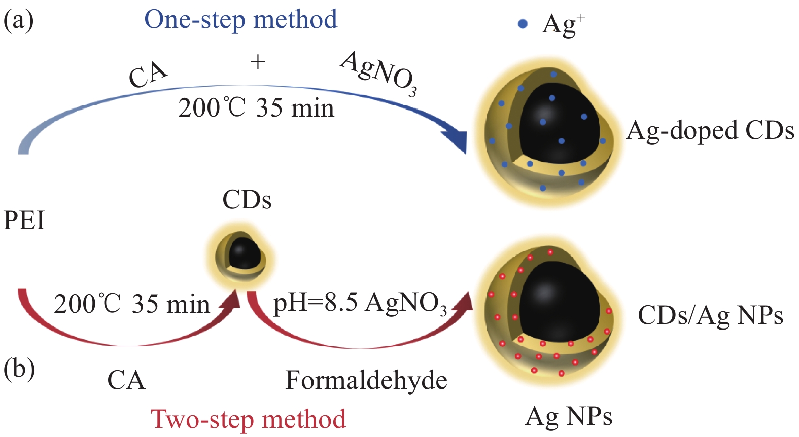
propertiesRef. Metal doping Ag AgNO3 CA, PEI Ag-
doped CDs1.8 +25.0 E. coli MIC: 50 μg·mL−1 [66] S. aureus MIC: 15 μg·mL−1 Cannabis sativa Ag@CDs — — E. coli MIC: 42 μg·mL−1 [67] S. aureus MIC: 42 μg·mL−1 Dopamine, cysteine NSCDAg — −44.0 E. coli MIC: 8 μg·mL−1 [68] NSCDAgAc — −29.0 E. coli MIC: 8 μg·mL−1 Ce Ce(NO3)3 CA Ce-CNDs 2-4 — S. aureus MIC: 200 μg·mL−1 [79] Cu Cu(CH3COO)2·H2O Tea Cu-CDs 0.9 — S. aureus MIC: 156 μg·mL−1 [80] Zn (CH₃COO)₂Zn CA, EDA Zn-CDs 1.8 — S. aureus SR: 89%
(Blue light: 40 min)[81] Non-metal doping N DETA Glucose NCQDs 5 +23.0 S. aureus DIZ: 15.5 mm [72] MRSA DIZ: 14.5 mm Polyvinylpyrrolidone N-CQDs 6.5 −6.5 E. coli MIC: 32 μg·mL−1 [74] PVAm CA CA∶ PVAm C-dots 12.6 +29.0 E. coli MIC: 1.56 mg·mL−1 [75] S. aureus MIC: 1.56 mg·mL−1 B. subtilis MIC: 0.75 mg·mL−1 Melamine, EDTA g-CNQDs 2 −36.0 E. coli SR: 99%
(100 μg·mL−1)[76] S. aureus SR: 90%
(100 μg·mL−1)Urea Glucose NGCD 5.6 −3.8 E. coli MIC: 19 μg·mL−1 [77] B H3BO3 Glucose BGCD 6.2 −18.0 E. coli MIC: 156 μg·mL−1 [77] S Poly (sodium-4-styrene sulfonate) S-CQDs 6.5 −47.2 E. coli MIC: 32 μg·mL−1 [74] (NH4)2S2O8 Glucose S-CDs 6.9 −5.5 E. coli MIC: 156 μg·mL−1 [77] Na2S2O8 Turmeric SGCD 8.5 +4.5 E. coli DIZ: 14 mm [83] P H3PO4 m-Aminophenol P-
doped CQDs3.4 +23.1 E. coli MIC: 1.23 mg·mL−1 [84] S. aureus MIC: 1.44 mg·mL−1 Co-doping N, S Urea, thiourea CA N, S-
doped CNDs— — E. coli SR: 68%
(500 μg·mL−1)[11] Ag,
NAgNO3, NH3∙H2O CA Ag, N-CQDs 5~9 — E. coli MIC: 250 μg·mL−1 [85] S. aureus MIC: 200 μg·mL−1 Ag,
SMPA,
AgNO3CA Ag@S-GQDs 28 — S. aureus MIC: 35 μg·mL−1 [86] Notes:DETA—Diethylenetriamine; PVAm—Polyethyleneamine; EDTA—Ethylene diamine tetraacetic acid; MPA—3-mercaptopropionic acid; SR—Sterilization rate; DIZ—Diameters of inhibition zone; NSCDs—Heteroatom (N and S) doped CDs; CNDs—Carbon nanodots; CQDs—Carbon quantum dots; GCD—Glucose CDs; GQD—Graphene quantum dots.
下载: 导出CSV
表 3 CDs复合材料的抗菌性能
Table 3. Antibacterial properties of CDs composites
Classification Antimicrobial Composite MIC/(μg·mL−1); SR/%; DIZ/mm Ref. E. coli S. aureus CDs/antibiotic RUT CDs-RUT 180 μg·mL−1 (Dark) 170 μg·mL−1 (Dark) [105] 150 μg·mL−1 (Light) 100 μg·mL−1 (Light) CDs/native compound CS N-doped C-dot (CS) 90.00% (100 μL) 92.00% (100 μL) [109] N, S doped C-dot (CS) 41.53% (100 μL) 48.54% (100 μL) Cur CQDs/Cur — >99.9% [110] CDs/inorganic compound CDs/Ag NPs Ag NPs CDs/Ag NPs 20 μg·mL−1 5 μg·mL−1 [66] Ag NPs/CDs 40 μg·mL−1 20 μg·mL−1 [90] ACMCD-Ag 10 μg·mL−1 5 μg·mL−1 [92] CDs/Ag NPs 12-13 mm 13-15 mm [93] CQDs/Ag NPs 10.88 mm 11.22 mm [94] Ag NPs-ACNPs 63.64% 93.49% [95] Ag-GQDs — 25 μg·mL−1 [96] CDs/metallic oxide ZnO ZnO@CQDs 6 mg·mL−1 8 mg·mL−1 [102] CDs/peroxide H2O2 H2O2/CDs 88%
(0.59 mM H2O2/8 μg·mL−1 CDs)— [59] CDs/Fe Fe Fe-CDs 99.85% 99.68% [106] CDs/organic compound CDs/dye MB CDs/MB 100%(1 μg·mL−1 MB/5 μg·mL−1 CDs) — [107] TB CDs/TB 100%(1 μg·mL−1 TB/
5 μg·mL−1 CDs)— BODIPY BODIPY@n-CDs — 256 μg·mL−1 [108] BODIPY@p-CDs — 128 μg·mL−1 CDs/polymer PVA N-doped C-dot (PVA) 47.53% (100 μL) 36.49% (100 μL) [109] N, S doped C-dot (PVA) 56.47% (100 μL) 46.51% (100 μL) PS N-doped C-dot (PS) 70.50% (100 μL) 39.86% (100 μL) N, S doped C-dot (PS) 61.65% (100 μL) 39.41% (100 μL) Notes:RUT—Rutin; CS—Chitosan; Cur—Curcumin; MB—Methylene blue; TB—Toluidine blue; BODIPY—Fluoroborondipyrrole; PVA—Polyvinyl alcohol; PS—Polysulfonatestyrene; ACMCD—Amphiphilic cow milk-derived CDs; ACNPs—Activated carbon nanoparticle.
下载: 导出CSV
-
[1] WILLYARD C. The drug-resistant bacteria that pose the greatest health threats[J]. Nature,2017,543:15. doi: 10.1038/nature.2017.21550 [2] WANG Y, YANG Y, SHI Y, et al. Antibiotic-free antibacterial strategies enabled by nanomaterials: Progress and perspectives[J]. Advanced Materials,2020,32(18):1904106. doi: 10.1002/adma.201904106 [3] ZHEN N, WANG X Y, LI X, et al. Protein-based natural antibacterial materials and their applications in food preservation[J]. Microbial Biotechnology,2022,15(5):1324-1338. doi: 10.1111/1751-7915.13918 [4] 莫尊理, 胡惹惹, 王雅雯, 等. 抗菌材料及其抗菌机理[J]. 材料导报, 2014, 28(1):50-52, 90.MO Zunli, HU Rere, WANG Yawen, et al. Review of antibacterial materials and their mechanisms[J]. Materials Reports,2014,28(1):50-52, 90(in Chinese). [5] 王静, 水中和, 冀志江, 等. 银系无机抗菌材料研究进展[J]. 材料导报, 2013, 27(17):59-64, 78. doi: 10.3969/j.issn.1005-023X.2013.17.012WANG Jing, SHUI Zhonghe, JI Zhijiang, et al. Research progress of the silver-typed inorganic antibacterial materials[J]. Materials Reports,2013,27(17):59-64, 78(in Chinese). doi: 10.3969/j.issn.1005-023X.2013.17.012 [6] 李媛, 韩玲珏, 王玥, 等. 光催化抗菌剂在医用抗菌方面的应用进展[J]. 中国材料进展, 2023, 42(2): 144-154.LI Yuan, HAN Lingjue, WANG Yue, et al. Application progress of photocatalytic antibacterial agents in medical antibacterial[J]. Materials China, 2023, 42(2): 144-154(in Chinese). [7] 姚希燕, 唐晓宁, 王晓楠, 等. 无机抗菌材料抗菌机理研究进展[J]. 材料导报, 2021, 35(1):1105-1111. doi: 10.11896/cldb.19090190YAO Xiyan, TANG Xiaoning, WANG Xiaonan, et al. Research progress on antibacterial mechanisms of inorganic antibacterial materials[J]. Materials Reports,2021,35(1):1105-1111(in Chinese). doi: 10.11896/cldb.19090190 [8] LI P F, SUN L, XUE S S, et al. Recent advances of carbon dots as new antimicrobial agents[J]. SmartMat,2022,3(2):226-248. doi: 10.1002/smm2.1131 [9] LI S, LI L, TU H Y, et al. The development of carbon dots: From the perspective of materials chemistry[J]. Materials Today,2021,51:188-207. doi: 10.1016/j.mattod.2021.07.028 [10] TRUSKEWYCZ A, YIN H, HALBERG N, et al. Carbon dot therapeutic platforms: Administration, distribution, metabolism, excretion, toxicity, and therapeutic potential[J]. Small, 2022, 18(16): 2106342. [11] CHATZIMITAKOS T G, KASOUNI A I, TROGANIS A N, et al. Exploring the antibacterial potential and unraveling the mechanism of action of non-doped and heteroatom-doped carbon nanodots[J]. Journal of Nanoparticle Research,2020,22(2):1-13. [12] YAN F, JIANG Y, SUN X, et al. Surface modification and chemical functionalization of carbon dots: A review[J]. Microchimica Acta,2018,185(9):1-34. [13] JOHN V L, NAIR Y, VINOD T P. Doping and surface modification of carbon quantum dots for enhanced functionalities and related applications[J]. Particle & Particle Systems Characterization,2021,38(11):2100170. [14] DOLMANS D E, FUKUMURA D, JAIN R K. Photodynamic therapy for cancer[J]. Nature Reviews Cancer,2003,3(5):380-387. doi: 10.1038/nrc1071 [15] HU X Q, HUANG Y Y, WANG Y G, et al. Antimicrobial photodynamic therapy to control clinically relevant biofilm infections[J]. Frontiers in Microbiology,2018,9:1299. doi: 10.3389/fmicb.2018.01299 [16] KIM A, ZHOU J, SAMADDAR S, et al. An implantable ultrasonically-powered micro-light-source (µlight) for photodynamic therapy[J]. Scientific Reports,2019,9(1):1-9. doi: 10.1038/s41598-019-38554-2 [17] NAZZAL S, CHEN C P, TSAI T. Nanotechnology in antimicrobial photodynamic inactivation[J]. Journal of Food and Drug Analysis,2011,19(4):12. [18] WU X, ABBAS K, YANG Y, et al. Photodynamic anti-bacteria by carbon dots and their nano-composites[J]. Pharmaceuticals,2022,15(4):487. doi: 10.3390/ph15040487 [19] GAO Z, YANG D Z, WAN Y, et al. One-step synthesis of carbon dots for selective bacterial inactivation and bacterial differentiation[J]. Analytical and Bioanalytical Chemistry,2020,412:871-880. doi: 10.1007/s00216-019-02293-0 [20] DONG X L, LIANG W X, MEZIANI M J, et al. Carbon dots as potent antimicrobial agents[J]. Theranostics,2020,10(2):671. doi: 10.7150/thno.39863 [21] YU J K, YONG X, TANG Z Y, et al. Theoretical understanding of structure-property relationships in luminescence of carbon dots[J]. The Journal of Physical Chemistry Letters,2021,12(32):7671-7687. doi: 10.1021/acs.jpclett.1c01856 [22] JIA Q Y, SONG Q, LI P, et al. Rejuvenated photodynamic therapy for bacterial infections[J]. Advanced Healthcare Materials,2019,8(14):1900608. doi: 10.1002/adhm.201900608 [23] ZHAI Y, ZHANG B, SHI R, et al. Carbon dots as new building blocks for electrochemical energy storage and electrocatalysis[J]. Advanced Energy Materials,2022,12(6):2103426. doi: 10.1002/aenm.202103426 [24] SONG Y B, ZHU S J, YANG B. Bioimaging based on fluorescent carbon dots[J]. RSC Advances,2014,4(52):27184-27200. doi: 10.1039/c3ra47994c [25] 陈童, 刘兴华, 郑静霞, 等. 碳点基白光荧光薄膜的研究进展[J]. 复合材料学报, 2022, 39(1):48-63.CHEN Tong, LIU Xinghua, ZHENG Jingxia, et al. Research progress of carbon dots based white light emitting fluorescent films[J]. Acta Materiae Compositae Sinica,2022,39(1):48-63(in Chinese). [26] XIA C L, ZHU S J, FENG T L, et al. Evolution and synthesis of carbon dots: From carbon dots to carbonized polymer dots[J]. Advanced Science,2019,6(23):1901316. doi: 10.1002/advs.201901316 [27] LI P L, YANG X, ZHANG X H, et al. Surface chemistry-dependent antibacterial and antibiofilm activities of polyamine-functionalized carbon quantum dots[J]. Journal of Materials Science,2020,55(35):16744-16757. doi: 10.1007/s10853-020-05262-6 [28] GHIRARDELLO M, RAMOS-SORIANO J, GALAN M C. Carbon dots as an emergent class of antimicrobial agents[J]. Nanomaterials,2021,11(8):1877. doi: 10.3390/nano11081877 [29] 刘晨艳, 闫凯, 马建中. 碳点及其纳米复合材料抗菌性能的研究进展[J]. 化工新型材料, 2022, 50(1):56-61.LIU Chenyan, YAN Kai, MA Jianzhong. Research progress on antibacterial property of carbon quantum dots and their nanocomposite[J]. New Chemical Materials,2022,50(1):56-61(in Chinese). [30] 张超, 张利, 刘兴华, 等. 碳纳米材料的抗菌性及在生物医学中的应用研究进展[J]. 材料导报, 2020, 34(S1):53-57.ZHANG Chao, ZHANG Li, LIU Xinghua, et al. Research advances in antibacterial properties and applications in biomedicine of carbon nanomaterials[J]. Materials Reports,2020,34(S1):53-57(in Chinese). [31] CHEN S, H GUO H X, CUI M, et al. Interaction of particles with mucosae and cell membranes[J]. Colloids and Surfaces B-Biointerfaces,2020,186:110657. doi: 10.1016/j.colsurfb.2019.110657 [32] JIAN H J, WU R S, LIN T Y, et al. Super-cationic carbon quantum dots synthesized from spermidine as an eye drop formulation for topical treatment of bacterial keratitis[J]. ACS Nano,2017,11(7):6703-6716. doi: 10.1021/acsnano.7b01023 [33] JOSHI A S, SINGH P, MIJAKOVIC I. Interactions of gold and silver nanoparticles with bacterial biofilms: Molecular interactions behind inhibition and resistance[J]. International Journal of Molecular Sciences,2020,21(20):7658. doi: 10.3390/ijms21207658 [34] LINKLATER D P, BAULIN V A, LE GUÉVEL X, et al. Antibacterial action of nanoparticles by lethal stretching of bacterial cell membranes[J]. Advanced Materials,2020,32(52):2005679. doi: 10.1002/adma.202005679 [35] 梁春燕. 碳点的可控制备及其与DNA/蛋白质的相互作用[D]. 黄石: 湖北师范大学, 2019.LIANG Chunyan. Controllable preparation of carbon dots and their interaction with DNA and protein[D]. Huangshi: Hubei Normal University, 2019(in Chinese). [36] PAN T, CHEN H H, GAO X, et al. Engineering efficient artificial nanozyme based on chitosan grafted Fe-doped-carbon dots for bacteria biofilm eradication[J]. Journal of Hazardous Materials,2022,435:128996. doi: 10.1016/j.jhazmat.2022.128996 [37] LIU M, HUANG L, XU X Y, et al. Copper doped carbon dots for addressing bacterial biofilm formation, wound infection, and tooth staining[J]. ACS Nano,2022,16(6):9479-9497. doi: 10.1021/acsnano.2c02518 [38] RISTIC B Z, MILENKOVIC M M, DAKIC I R, et al. Photodynamic antibacterial effect of graphene quantum dots[J]. Biomaterials,2014,35(15):4428-4435. doi: 10.1016/j.biomaterials.2014.02.014 [39] QIE X W, ZAN M H, GUI P, et al. Design, synthesis, and application of carbon dots with synergistic antibacterial activity[J]. Frontiers in Bioengineering and Biotechnology, 2022, 10: 894100. [40] WEN F Z, LI P Y, MENG H R, et al. Nitrogen-doped carbon dots/curcumin nanocomposite for combined photodynamic/photothermal dual-mode antibacterial therapy[J]. Photodiagnosis and Photodynamic Therapy,2022,39:103033. doi: 10.1016/j.pdpdt.2022.103033 [41] PRAJAPATI J D, KLEINEKATHÖFER U, WINTERHALTER M. How to enter a bacterium: bacterial porins and the permeation of antibiotics[J]. Chemical Reviews,2021,121(9):5158-5192. doi: 10.1021/acs.chemrev.0c01213 [42] VARGHESE M, BALACHANDRAN M. Antibacterial efficiency of carbon dots against gram-positive and gram-negative bacteria: A review[J]. Journal of Environmental Chemical Engineering,2021,9(6):106821. doi: 10.1016/j.jece.2021.106821 [43] 张荣. 新型荧光碳点的制备及其在传感、生物成像和抗菌中的应用[D]. 太原: 山西医科大学, 2019.ZHANG Rong. Preparation of novel fluorescent carbon dots and their application in sensing, bioimaging and antibacterum[D]. Taiyuan: Shanxi Medical University, 2019(in Chinese). [44] LU F, MA Y R, WANG H B, et al. Water-solvable carbon dots derived from curcumin and citric acid with enhanced broad-spectrum antibacterial and antibiofilm activity[J]. Materials Today Communications,2021,26:102000. doi: 10.1016/j.mtcomm.2020.102000 [45] LI J C, MAO H L, KAWAZOE N, et al. Insight into the interactions between nanoparticles and cells[J]. Biomaterials Science,2017,5(2):173-189. doi: 10.1039/C6BM00714G [46] SHANG L, NIENHAUS K, NIENHAUS G U. Engineered nanoparticles interacting with cells: Size matters[J]. Journal of Nanobiotechnology,2014,12(1):1-11. doi: 10.1186/1477-3155-12-5 [47] ZHAO F, ZHAO Y, LIU Y, et al. Cellular uptake, intracellular trafficking, and cytotoxicity of nanomaterials[J]. Small,2011,7(10):1322-1337. doi: 10.1002/smll.201100001 [48] SUN B H, WU F, ZHANG Q C. Insight into the effect of particle size distribution differences on the antibacterial activity of carbon dots[J]. Journal of Colloid and Interface Science,2021,584:505-519. doi: 10.1016/j.jcis.2020.10.015 [49] DI LORENZO F, DUDA K A, LANZETTA R, et al. A journey from structure to function of bacterial lipopolysaccharides[J]. Chemical Reviews,2021,122(20):15767-15821. [50] WANG H B, LU F, MA C Q, et al. Carbon dots with positive surface charge from tartaric acid and m-aminophenol for selective killing of gram-positive bacteria[J]. Journal of Materials Chemistry B,2021,9(1):125-130. doi: 10.1039/D0TB02332A [51] BING W, SUN H J, YAN Z Q, et al. Programmed bacteria death induced by carbon dots with different surface charge[J]. Small,2016,12(34):4713-4718. doi: 10.1002/smll.201600294 [52] WU L, GAO Y R, ZHAO C F, et al. Synthesis of curcumin-quaternized carbon quantum dots with enhanced broad-spectrum antibacterial activity for promoting infected wound healing[J]. Biomaterials Advances,2022,133:112608. doi: 10.1016/j.msec.2021.112608 [53] MAHAT N A, NOR N S M, SHAMSUDIN S A. Effects of positive carbon quantum dots on gram-negative bacteria as an antimicrobial agent[J]. Journal of Inorganic and Organometallic Polymers and Materials, 2022, 32, 2428-2440. [54] ZHAO C F, WANG X W, YU L Y, et al. Quaternized carbon quantum dots with broad-spectrum antibacterial activity for the treatment of wounds infected with mixed bacteria[J]. Acta Biomaterialia,2022,138:528-544. doi: 10.1016/j.actbio.2021.11.010 [55] SVIRIDOVA E, BARRAS A, ADDAD A, et al. Surface modification of carbon dots with tetraalkylammonium moieties for fine tuning their antibacterial activity[J]. Materials Science and Engineering: C, 2022, 134: 112697. [56] LI Y J, HARROUN S G, SU Y C, et al. Synthesis of self-assembled spermidine-carbon quantum dots effective against multidrug-resistant bacteria[J]. Advanced Healthcare Materials,2016,5(19):2545-2554. doi: 10.1002/adhm.201600297 [57] GAGIC M, KOCIOVA S, SMERKOVA K, et al. One-pot synthesis of natural amine-modified biocompatible carbon quantum dots with antibacterial activity[J]. Journal of Colloid and Interface Science,2020,580:30-48. doi: 10.1016/j.jcis.2020.06.125 [58] DEVKOTA A, PANDEY A, YADEGARI Z, et al. Amine-coated carbon dots (NH2-FCDs) as novel antimicrobial agent for gram-negative bacteria[J]. Frontiers in Nanotechnology, 2021, 3: 78. [59] DONG X L, AWAK M A, TOMLINSON N, et al. Antibacterial effects of carbon dots in combination with other antimicrobial reagents[J]. PloS One,2017,12(9):e0185324. doi: 10.1371/journal.pone.0185324 [60] OTIS G, BHATTACHARYA S, MALKA O, et al. Selective labeling and growth inhibition of Pseudomonas aeruginosa by aminoguanidine carbon dots[J]. ACS Infectious Diseases,2018,5(2):292-302. [61] YANG J, ZHANG X, MA Y H, et al. Carbon dot-based platform for simultaneous bacterial distinguishment and antibacterial applications[J]. ACS Applied Materials & Interfaces,2016,8(47):32170-32181. [62] JIJIE R, BARRAS A, BOUCKAERT J, et al. Enhanced antibacterial activity of carbon dots functionalized with ampicillin combined with visible light triggered photodynamic effects[J]. Colloids and Surfaces B: Biointerfaces,2018,170:347-354. doi: 10.1016/j.colsurfb.2018.06.040 [63] PARK Y, YOO J, LIM B, et al. Improving the functionality of carbon nanodots: Doping and surface functionalization[J]. Journal of Materials Chemistry A,2016,4(30):11582-11603. doi: 10.1039/C6TA04813G [64] MIAO S H, LIANG K, ZHU J J, et al. Hetero-atom-doped carbon dots: Doping strategies, properties and applications[J]. Nano Today,2020,33:100879. doi: 10.1016/j.nantod.2020.100879 [65] JAIN J, ARORA S, RAJWADE J M, et al. Silver nanoparticles in therapeutics: Development of an antimicrobial gel formulation for topical use[J]. Molecular Pharmaceutics,2009,6(5):1388-1401. doi: 10.1021/mp900056g [66] ZHAO D, LIU X M, ZHANG R, et al. Preparation of two types of silver-doped fluorescent carbon dots and determination of their antibacterial properties[J]. Journal of Inorganic Biochemistry,2021,214:111306. doi: 10.1016/j.jinorgbio.2020.111306 [67] RAINA S, THAKUR A, SHARMA A, et al. Bactericidal activity of Cannabis sativa phytochemicals from leaf extract and their derived carbon dots and Ag@carbon dots[J]. Materials Letters,2020,262:127122. doi: 10.1016/j.matlet.2019.127122 [68] JANA J, GAURI S S, GANGULY M, et al. Silver nanoparticle anchored carbon dots for improved sensing, catalytic and intriguing antimicrobial activity[J]. Dalton Transactions,2015,44(47):20692-20707. doi: 10.1039/C5DT03858H [69] WANG M Q, SU Y T, LIU Y H, et al. Antibacterial fluorescent nano-sized lanthanum-doped carbon quantum dot embedded polyvinyl alcohol for accelerated wound healing[J]. Journal of Colloid and Interface Science,2022,608:973-983. doi: 10.1016/j.jcis.2021.10.018 [70] 苏玉天. 新型碳量子点的制备与性能研究[D]. 南京: 南京师范大学, 2017.SU Yutian. Study on the preparation and performance of new carbon quantum dots[D]. Nanjing: Nanjing Normal University, 2017(in Chinese). [71] ZHANG M, WANG W T, YUAN P, et al. Synthesis of lanthanum doped carbon dots for detection of mercury ion, multi-color imaging of cells and tissue, and bacteriostasis[J]. Chemical Engineering Journal,2017,330:1137-1147. doi: 10.1016/j.cej.2017.07.166 [72] ZHAO C F, WANG X W, WU L, et al. Nitrogen-doped carbon quantum dots as an antimicrobial agent against staphylococcus for the treatment of infected wounds[J]. Colloids and Surfaces B-Biointerfaces,2019,179:17-27. doi: 10.1016/j.colsurfb.2019.03.042 [73] WANG H, SONG Z, GU J, et al. Nitrogen-doped carbon quantum dots for preventing biofilm formation and eradicating drug-resistant bacteria infection[J]. ACS Biomaterials Science & Engineering,2019,5(9):4739-4749. [74] TRAVLOU N A, GIANNAKOUDAKIS D A, ALGARRA M, et al. S-and N-doped carbon quantum dots: Surface chemistry dependent antibacterial activity[J]. Carbon,2018,135:104-111. doi: 10.1016/j.carbon.2018.04.018 [75] SUTEKIN S D, SAHINER M, SUNER S S, et al. Poly(vinylamine) derived N-doped C-dots with antimicrobial and antibiofilm activities[J]. Journal of Carbon Research,2021,7(2):40. doi: 10.3390/c7020040 [76] YADAV P, NISHANTHI S T, PUROHIT B, et al. Metal-free visible light photocatalytic carbon nitride quantum dots as efficient antibacterial agents: an insight study[J]. Carbon,2019,152:587-597. doi: 10.1016/j.carbon.2019.06.045 [77] EZATI P, RHIM J W, MOLAEI R, et al. Preparation and characterization of B, S, and N-doped glucose carbon dots: Antibacterial, antifungal, and antioxidant activity[J]. Sustainable Materials and Technologies,2022,32:e00397. doi: 10.1016/j.susmat.2022.e00397 [78] KNOBLAUCH R, HARVEY A, RA E, et al. Antimicrobial carbon nanodots: Photodynamic inactivation and dark antimicrobial effects on bacteria by brominated carbon nanodots[J]. Nanoscale,2021,13(1):85-99. doi: 10.1039/D0NR06842J [79] ZHANG M Z, ZHAI X Y, MA T F, et al. Multifunctional cerium doped carbon dots nanoplatform and its applications for wound healing[J]. Chemical Engineering Journal,2021,423:130301. doi: 10.1016/j.cej.2021.130301 [80] QING W X, CHEN K, YANG Y Y, et al. Cu2+-doped carbon dots as fluorescence probe for specific recognition of Cr(VI) and its antimicrobial activity[J]. Microchemical Journal,2020,152:104262. doi: 10.1016/j.microc.2019.104262 [81] LIU D N, YANG M X, LIU X C, et al. Zinc-doped carbon dots as effective blue-light-activated antibacterial agent[J]. Nano,2021,16(3):2150031. doi: 10.1142/S1793292021500314 [82] ZHONG Z L, ZHANG Y Y, FU X Y, et al. Construction of photo-induced zinc-doped carbon dots based on drug-resistant bactericides and their application for local treatment[J]. Nanoscale Advances, 2022, 4(24): 5365-5377. [83] ROY S, EZATI P, RHIM J W, et al. Preparation of turmeric-derived sulfur-functionalized carbon dots: Antibacterial and antioxidant activity[J]. Journal of Materials Science,2022,57(4):2941-2952. doi: 10.1007/s10853-021-06804-2 [84] CHAI S Q, ZHOU L J, PEI S C, et al. P-doped carbon quantum dots with antibacterial activity[J]. Micromachines,2021,12(9):1116. doi: 10.3390/mi12091116 [85] WANG J, ZHU Y X, XIE X F, et al. Effect of ultra-trace Ag doping on the antibacterial performance of carbon quantum dots[J]. Journal of Environmental Chemical Engineering,2022,10(2):107112. doi: 10.1016/j.jece.2021.107112 [86] KADIAN S, MANIK G, DAS N, et al. Synthesis, characterization and investigation of synergistic antibacterial activity and cell viability of silver-sulfur doped graphene quantum dot (Ag@S-GQDs) nanocomposites[J]. Journal of Materials Chemistry B,2020,8(15):3028-3037. doi: 10.1039/C9TB02823D [87] LIU W J, GU H, LIU W K, et al. NIR-emitting carbon dots for discriminative imaging and photo-inactivation of pathogenic bacteria[J]. Chemical Engineering Journal,2022,450:137384. doi: 10.1016/j.cej.2022.137384 [88] LE OUAY B, STELLACCI F. Antibacterial activity of silver nanoparticles: A surface science insight[J]. Nano Today,2015,10(3):339-354. doi: 10.1016/j.nantod.2015.04.002 [89] MA J L, LI K X, GU S B, et al. Antimicrobial carbon-dot-stabilized silver nanoparticles[J]. New Journal of Chemistry,2022,46(5):2546-2552. doi: 10.1039/D1NJ05798G [90] WANG P, Y SONG Y Z, MEI Q, et al. Sliver nanoparticles@ carbon dots for synergistic antibacterial activity[J]. Applied Surface Science,2022,600:154125. doi: 10.1016/j.apsusc.2022.154125 [91] LIU T Y, PANG Q Y, MAI K, et al. Silver nanoparticle@carbon quantum dot composite as an antibacterial agent[J]. RSC Advances,2022,12(16):9621-9627. doi: 10.1039/D2RA00561A [92] HAN S, ZHANG H, XIE Y J, et al. Application of cow milk-derived carbon dots/AgNPs composite as the antibacterial agent[J]. Applied Surface Science,2015,328:368-373. doi: 10.1016/j.apsusc.2014.12.074 [93] WEI X J, CHENG F, YAO Y, et al. Facile synthesis of a carbon dots and silver nanoparticles (CDs/AgNPs) composite for antibacterial application[J]. RSC Advances,2021,11(30):18417-18422. doi: 10.1039/D1RA02600C [94] CHENG Y L, WEI Y, FANG C Q, et al. Facile synthesis of CQDs/AgNPs composites with photoluminescence and their potential application in antibacterial materials[J]. Inorganic Chemistry Communications,2021,134:109059. doi: 10.1016/j.inoche.2021.109059 [95] WIBAWA P J, NUR M, ASY’ARI M, et al. Green synthesized silver nanoparticles immobilized on activated carbon nanoparticles: Antibacterial activity enhancement study and its application on textiles fabrics[J]. Molecules,2021,26(13):3790. doi: 10.3390/molecules26133790 [96] HABIBA K, BRACHO-RINCON D P, GONZALEZ-FELICIANO J A, et al. Synergistic antibacterial activity of PEGylated silver-graphene quantum dots nanocomposites[J]. Applied Materials Today,2015,1(2):80-87. doi: 10.1016/j.apmt.2015.10.001 [97] DA SILVA B L, CAETANO B L, CHIARI-ANDRÉO B G, et al. Increased antibacterial activity of ZnO nanoparticles: Influence of size and surface modification[J]. Colloids and Surfaces B: Biointerfaces,2019,177:440-447. doi: 10.1016/j.colsurfb.2019.02.013 [98] MYDEEN S S, KUMAR R R, KOTTAISAMY M, et al. Biosynthesis of ZnO nanoparticles through extract from prosopis julifora plant leaf: Antibacterial activities and a new approach by rust-induced photocatalysis[J]. Journal of Saudi Chemical Society,2020,24(5):393-406. doi: 10.1016/j.jscs.2020.03.003 [99] QI K Z, CHENG B, YU J G, et al. Review on the improvement of the photocatalytic and antibacterial activities of ZnO[J]. Journal of Alloys and Compounds,2017,727:792-820. doi: 10.1016/j.jallcom.2017.08.142 [100] MYDEEN S S, KUMAR R R, SIVAKUMAR R, et al. Graphene quantum dots/ZnO nanocomposite: Synthesis, characterization, mechanistic investigations of photocatalytic and antibacterial activities[J]. Chemical Physics Letters,2020,761:138009. doi: 10.1016/j.cplett.2020.138009 [101] ABEBE B, ZEREFFA E A, TADESSE A, et al. A review on enhancing the antibacterial activity of ZnO: Mechanisms and microscopic investigation[J]. Nanoscale Research Letters,2020,15(1):1-19. doi: 10.1186/s11671-020-03418-6 [102] GAO D, ZHAO P, LYU B, et al. Carbon quantum dots decorated on ZnO nanoparticles: an efficient visible-light responsive antibacterial agents[J]. Applied Organometallic Chemistry,2020,34(8):e5665. [103] YAN Y Y, KUANG W C, SHI L J, et al. Carbon quantum dot-decorated TiO2 for fast and sustainable antibacterial properties under visible-light[J]. Journal of Alloys and Compounds,2019,777:234-243. doi: 10.1016/j.jallcom.2018.10.191 [104] THAKUR M, PANDEY S, MEWADA A, et al. Antibiotic conjugated fluorescent carbon dots as a theranostic agent for controlled drug release, bioimaging, and enhanced antimicrobial activity[J]. Journal of Drug Delivery,2014,2014:282193. [105] TEJWAN N, KUNDU M, GHOSH N, et al. Synthesis of green carbon dots as bioimaging agent and drug delivery system for enhanced antioxidant and antibacterial efficacy[J]. Inorganic Chemistry Communications,2022,139:109317. doi: 10.1016/j.inoche.2022.109317 [106] LIU Y H, XU B L, LU M Z, et al. Ultrasmall Fe-doped carbon dots nanozymes for photoenhanced antibacterial therapy and wound healing[J]. Bioactive Materials,2022,12:246-256. doi: 10.1016/j.bioactmat.2021.10.023 [107] DONG X L, BOND A E, PAN N, et al. Synergistic photoactivated antimicrobial effects of carbon dots combined with dye photosensitizers[J]. International Journal of Nanomedicine,2018,13:8025. doi: 10.2147/IJN.S183086 [108] MOU C J, WANG X Y, LIU Y C, et al. Positively charged BODIPY@carbon dot nanocomposites for enhanced photomicrobicidal efficacy and wound healing[J]. Journal of Materials Chemistry B,2022,10(39):8094-8099. doi: 10.1039/D2TB01539K [109] DANIEL S, KS S. Highly luminescent biocompatible doped nano carbon dot composites as efficient antibacterial agents[J]. Composite Interfaces,2021,28(11):1155-1170. doi: 10.1080/09276440.2020.1867466 [110] WEN F Z, LI P Y, ZHANG Y, et al. Preparation, characterization of green tea carbon quantum dots/curcumin antioxidant and antibacterial nanocomposites[J]. Journal of Molecular Structure, 2022, 1273: 134247. [111] EZATI P, RHIM J W, MOLAEI R, et al. Cellulose nanofiber-based coating film integrated with nitrogen-functionalized carbon dots for active packaging applications of fresh fruit[J]. Postharvest Biology and Technology,2022,186:111845. doi: 10.1016/j.postharvbio.2022.111845 [112] SUWAILEH W, PATHAK N, SHON H, et al. Forward osmosis membranes and processes: A comprehensive review of research trends and future outlook[J]. Desalination,2020,485:114455. doi: 10.1016/j.desal.2020.114455 [113] MAHAT N A, SHAMSUDIN S A, JULLOK N, et al. Carbon quantum dots embedded polysulfone membranes for antibacterial performance in the process of forward osmosis[J]. Desalination,2020,493:114618. doi: 10.1016/j.desal.2020.114618 [114] KAMOUN E A, KENAWY E R S, CHEN X. A review on polymeric hydrogel membranes for wound dressing applications: PVA-based hydrogel dressings[J]. Journal of Advanced Research,2017,8(3):217-233. doi: 10.1016/j.jare.2017.01.005 [115] YANG X, LI P L, TANG W T, et al. A facile injectable carbon dot/oxidative polysaccharide hydrogel with potent self-healing and high antibacterial activity[J]. Carbohydrate Polymers,2021,251:117040. doi: 10.1016/j.carbpol.2020.117040 [116] NIE X L, WU S L, MENSAH A, et al. Carbon quantum dots embedded electrospun nanofibers for efficient antibacterial photodynamic inactivation[J]. Materials Science & Engineering C—Materials for Biological Applications,2020,108:110377. [117] KOULIVAND H, SHAHBAZI A, VATANPOUR V, et al. Novel antifouling and antibacterial polyethersulfone membrane prepared by embedding nitrogen-doped carbon dots for efficient salt and dye rejection[J]. Materials Science & Engineering: C,2020,111:110787. -

 下载:
下载:


 点击查看大图

计量
- 文章访问数: 1183
- HTML全文浏览量: 386
- PDF下载量: 96
- 被引次数: 0
