


Evaluation method of biomaterial degradation based on in vivo imaging system
-
摘要: 可降解材料作为生物材料的重要组成部分,其体内降解性能的好坏往往决定着材料植入后的成败。因此,对材料体内降解的评价显得尤为重要。传统的生物材料体内降解评价方法需要在各取样点取出不同批次的降解样品,阻止了对同一个实验样品降解过程的连续测量,并且存在样品需求量大的问题。小动物活体成像系统(in vivo imaging system,IVIS)具有非侵入性、操作性强等特点,为解决上述问题提供了思路。本研究旨在建立一种利用小动物荧光成像系统检测可降解材料降解性能的方法,通过将近红外荧光染料经化学反应标记到可降解材料上,由荧光强度的变化反应材料的降解程度。体内降解实验表明此方法制得的荧光标记材料,荧光稳定性高,材料降解过程中荧光强度变化与质量损失拟合效果良好(R2=0.9994)。综上,该方法解决了测量材料降解样品量大的问题,并且提高了实验过程的连贯性。
-
关键词:
- 可降解医用高分子材料 /
- 体内降解 /
- 小动物活体成像系统 /
- 荧光标记 /
- 评价方法
Abstract: Degradable material is an important part of biomaterials, its degradation performance in vivo has great influence on the final success after implantation. Thus, the method of evaluating the degradation of the materials in vivo is crucial to the evaluation of material performance. The traditional method to estimate the degradation in vivo needs to take out different batches of degradation samples at each sampling point, which prevents the continuous measurement of the degradation process and it needs a large number of samples. In vivo imaging system (IVIS) has the characteristics of non-invasive and strong operability, which provides a way to solve the problems above. In this study, we established a method to detect the degradation performance of degradable materials with IVIS. The research method was that the near-infrared fluorescent dye was labeled on the degradable materials by chemical reaction, and then using the change of fluorescence intensity to reflect the degradation degree of the materials. The in vivo degradation experimental results show that the fluorescent labeling materials prepared by this method have high fluorescence stability, and the fitting effect of fluorescence intensity and mass loss in the process of material degradation is good (R2 = 0.9994). In conclusion, this method solves the problem of large amount of samples in the traditional method of measuring material degradation, and improves the continuity of the experimental process. -

图 1 聚乳酸-羟基乙酸共聚物(PLGA)、PLGA-叔丁氧羰基(Boc)及PLGA-NH2的FTIR图谱
Figure 1. FTIR spectra of poly(lactide-co-glycolide) acid (PLGA), PLGA-t-butyloxycarbonyl (Boc) and PLGA-NH2

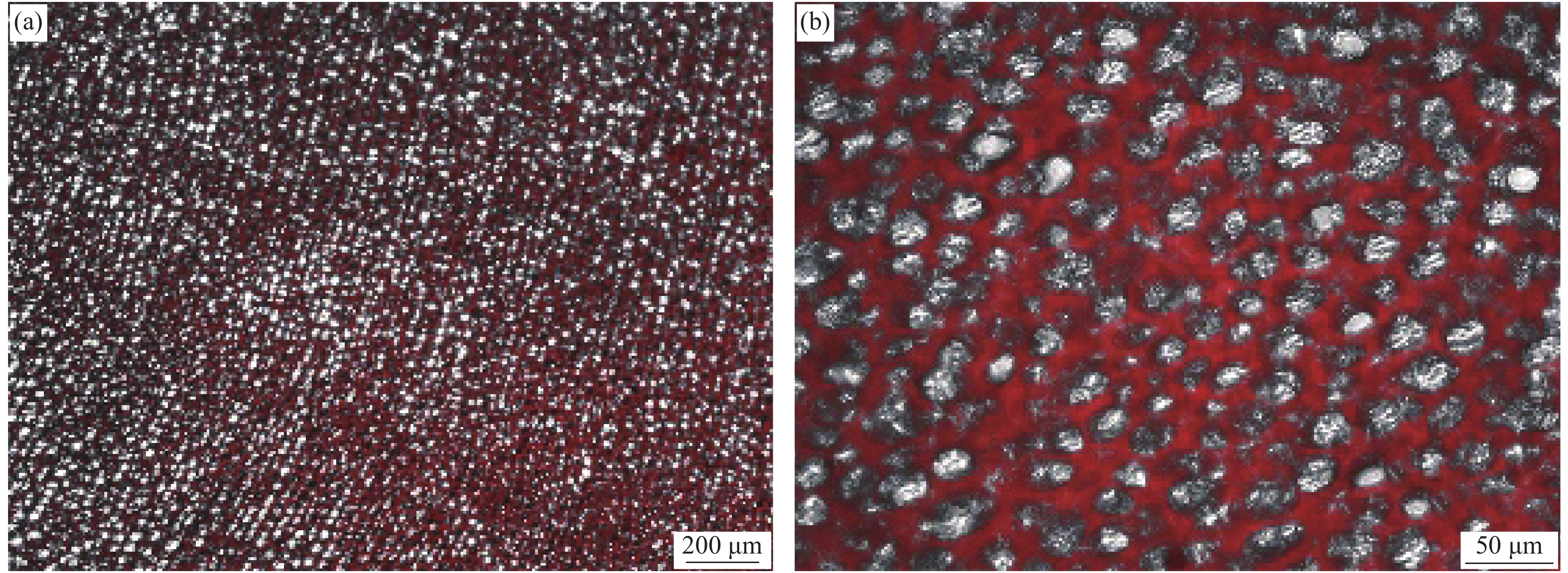
图 2 荧光显微镜拍摄的PLGA-cy5.5多孔膜的明场图像与暗场图像的叠加
Figure 2. Superposition of bright field image and dark field image of PLGA-cy5.5 porous membrane by fluorescence microscope

图 3 荧光显微镜拍摄的cy5.5标记的胶原蛋白海绵
Figure 3. cy5.5 labeled collagen sponge under the fluorescence microscope
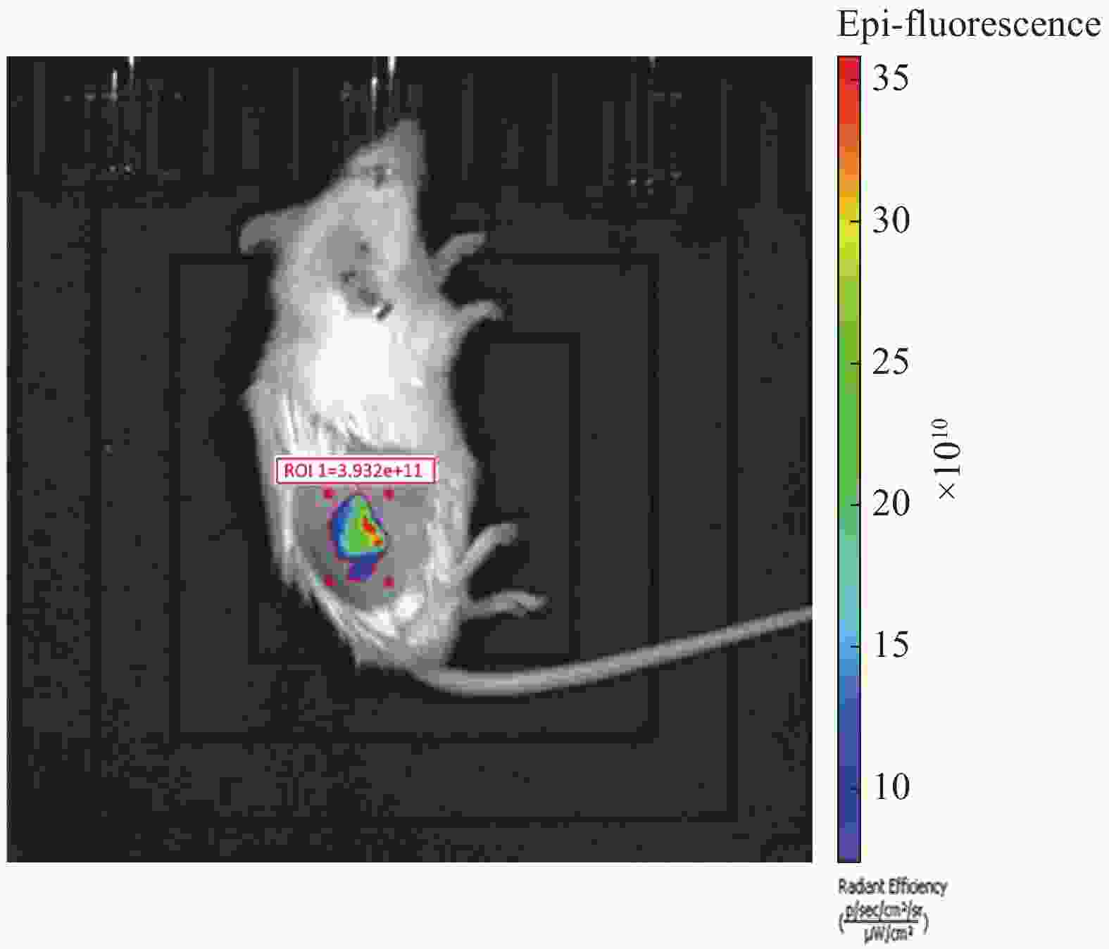
图 4 IVIS Lumina Ⅲ软件自动圈选感兴趣区域(ROI)
Figure 4. Automatically circle range of interest (ROI) in Living Image software

图 5 根据植入的PLGA-cy5.5多孔膜的实际大小、位置手动圈选ROI
Figure 5. Manually circle ROI according to actual size and location of implanted PLGA-cy5.5 porous membrane

图 6 分析cy5.5荧光标记的胶原蛋白海绵降解过程时使用相同大小与相同相对位置的选框圈定ROI
Figure 6. Using ROI of the same size and the same relative location to analyze the degradation process of the cy5.5 labeled collagen sponge

图 7 根据降解过程中cy5.5荧光标记的胶原蛋白海绵的实际大小和位置圈定ROI
Figure 7. Using ROI defined by the cy5.5 labeled collagen sponge’s actual size and location to analyze the degradation process

图 8 圈选小鼠无材料位置的皮肤作为背景荧光值(左侧)与植入cy5.5荧光标记的胶原蛋白海绵的荧光值相比较(右侧)
Figure 8. Skin without implanted material was selected as the background (left) and was compared to the fluorescent intensity of the implanted cy5.5 labeled collagen sponge (right)

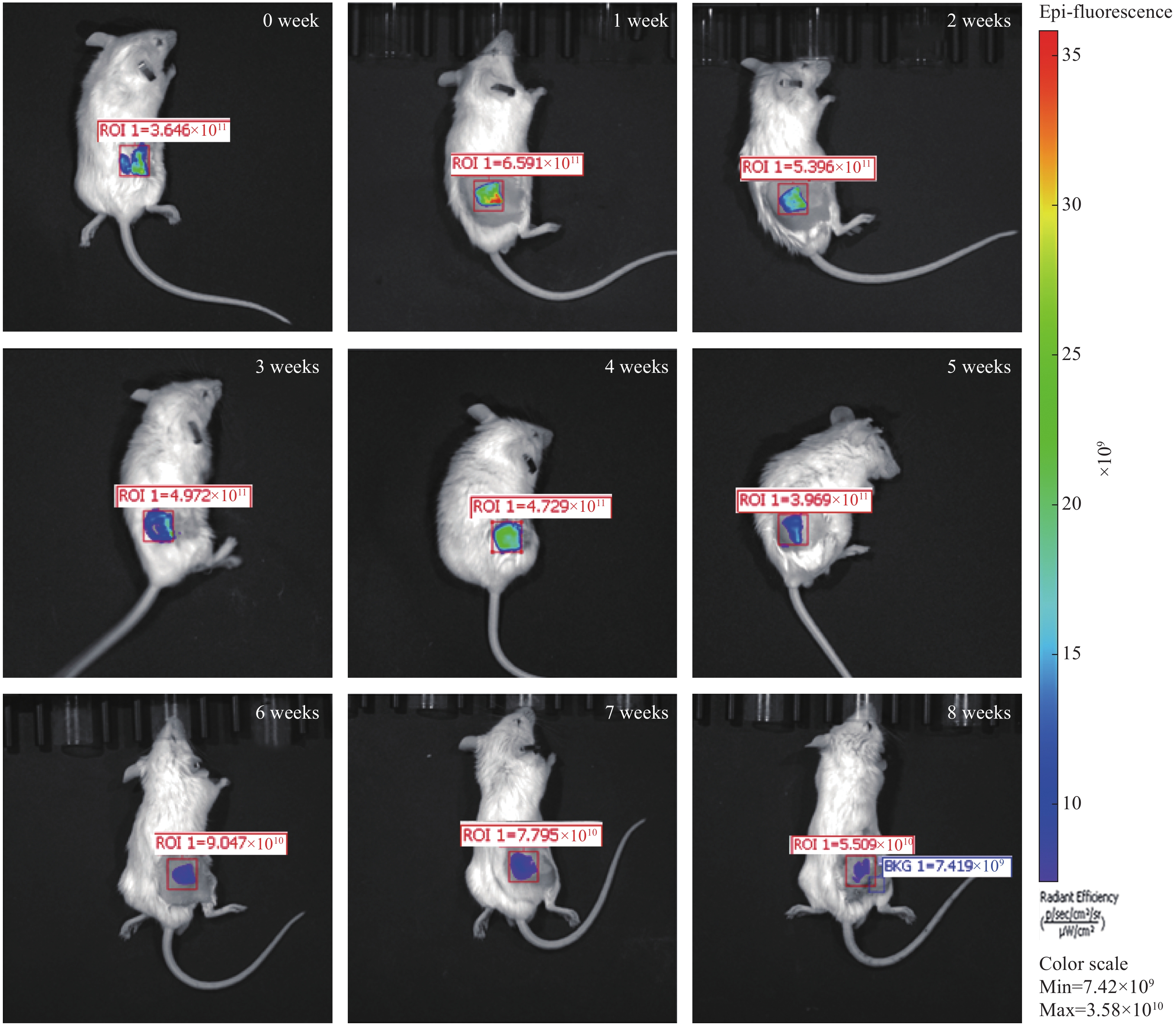
图 9 PLGA-cy5.5多孔膜在小鼠体内降解8周的荧光强度变化图
Figure 9. PLGA-cy5.5 porous membrane’s fluorescence intensity loss during 8 weeks degradation in rats
-
[1] HELLING A L, TSEKOURA E K, BIGGS M, et al. In vitro enzymatic degradation of tissue grafts and collagen biomaterials by matrix metalloproteinases: Improving the collagenase assay[J]. ACS Biomaterials Science & Engineering,2017,3(9):1922-1932. [2] CHU Z, ZHENG Q, GUO M, et al. The effect of fluid shear stress on the in vitro degradation of poly(lactide-co-glycolide) acid membranes[J]. Journal of Biomedical Materials Research Part A,2016,104(9):2315-2324. doi: 10.1002/jbm.a.35766 [3] LU L, PETER S J, LYMAN M D, et al. In vitro and in vivo degradation of porous poly(DL-lactic-co-glycolic acid) foams[J]. Biomaterials,2000,21(18):1837-1845. doi: 10.1016/S0142-9612(00)00047-8 [4] FREIER T, KUNZE C, NISCHAN C, et al. In vitro and in vivo degradation studies for development of a biodegradable patch based on poly(3-hydroxybutyrate)[J]. Biomaterials,2002,23(13):2649-2657. doi: 10.1016/S0142-9612(01)00405-7 [5] QIU L. In vivo degradation and tissue compatibility of polyphosphazene blend films[J]. Journal of Biomedical Engineering,2002,19(2):191-195. [6] BERGSMA J E, ROZEMA F R, BOS R R, et al. In vivo degradation and biocompatibility study of in vitro pre-degraded as-polymerized polyactide particles[J]. Biomaterials,1995,16(4):267-274. doi: 10.1016/0142-9612(95)93253-A [7] YANG F H, NIU X F, GU X N, et al. Biodegradable magnesium-incorporated poly(L-lactic acid) microspheres for manipulation of drug release and alleviation of inflammatory response[J]. ACS Applied Materials & Interfaces,2019,11(26):23546-23557. doi: 10.1021/acsami.9b03766 [8] NIU X F, LIU Z N, HU J, et al. Microspheres assembled from chitosan-graft-poly(lactic acid) micelle-like core-shell nanospheres for distinctly controlled release of hydrophobic and hydrophilic biomolecules[J]. Macromolecular Bioscience,2016,16:1039-1047. doi: 10.1002/mabi.201600020 [9] 潘永明, 金平, 徐剑钦, 等. 基于3.0 T磁共振成像系统初步观察WHBE兔脑部形态解剖结构[J]. 中国实验动物学报, 2017, 25(4):356-361.PAN Yongming, JIN Ping, XU Jianqin, et al. Preliminary observation of the anatomical structures of the brain in WHBE rabbits by 3.0 T magnetic resonance imaging system[J]. Acta Laboratorium Animalis Scientia Sinica,2017,25(4):356-361(in Chinese). [10] 李珂, 赵光, 高春芳, 等. 小动物活体成像技术的应用进展[J]. 实用医药杂志, 2012, 29(1):81-82. doi: 10.3969/j.issn.1671-4008.2012.01.057LI Ke, ZHAO Guang, GAO Chunfang, et al. Advances in the application of in vivo imaging technology[J]. Practical Journal of Medicine & Pharmacy,2012,29(1):81-82(in Chinese). doi: 10.3969/j.issn.1671-4008.2012.01.057 [11] 赵年欢, 崔邦平, 王朋, 等. 生物发光成像示踪干细胞移植的应用进展[J]. 巴楚医学, 2018, 1(1):125-128.ZHAO Nianhuan, CUI Bangping, WANG Peng, et al. Advances in the application of bioluminescence imaging tracing in stem cell transplantation[J]. Bachu Medical Journal,2018,1(1):125-128(in Chinese). [12] GONDI C S, VEERAVALLI K K, GORANTLA B, et al. Human umbilical cord blood stem cells show PDGF-D-dependent glioma cell tropism in vitro and in vivo[J]. Neuro Oncology,2010,12(5):453-465. [13] 柴凡, 周耘裔, 肖庚富. 活体生物发光成像技术及其在病毒感染研究中的应用[J]. 微生物学报, 2011, 51(4):431-437.CHAI Fan, ZHOU Yunyi, XIAO Gengfu. Bioluminescence in-vivo imaging technology and its application in the study of viral infection-A review[J]. Acta Microbiologica Sinica,2011,51(4):431-437(in Chinese). [14] GUTOWSKI M B, WILSON L, VAN GELDER R N, et al. In vivo bioluminescence imaging for longitudinal monitoring of inflammation in animal models of uveitis[J]. Investigative Ophthalmology & Visual Science,2017,58(3):1521-1528. [15] YANAGIHARA K, TAKIGAHIRA M, TAKESHITA F, et al. A photon counting technique for quantitatively evaluating progression of peritoneal tumor dissemination[J]. Cancer Research,2006,66(15):7532-7539. doi: 10.1158/0008-5472.CAN-05-3259 [16] IYER M, SALAZAR F B, WU L, et al. Bioluminescence imaging of systemic tumor targeting using a prostate-specific lentiviral vector[J]. Human Gene Therapy,2006,17(1):125-132. doi: 10.1089/hum.2006.17.125 [17] MA T C, HOU Y, ZENG J F, et al. Dual-ratiometric target-triggered fluorescent probe for simultaneous quantitative visualization of tumor microenvironment protease activity and pH & ITin vivo & IT[J]. Journal of the American Chemical Society,2018,140(1):211-218. doi: 10.1021/jacs.7b08900 [18] CHOI H S, GIBBS S L, LEE J H, et al. Targeted zwitterionic near-infrared fluorophores for improved optical imaging[J]. Nature Biotechnology,2013,31(2):148-153. doi: 10.1038/nbt.2468 [19] CHOI K Y, CHUNG H, MIN K H, et al. Self-assembled hyaluronic acid nanoparticles for active tumor targeting[J]. Biomaterials,2010,31(1):106-114. doi: 10.1016/j.biomaterials.2009.09.030 [20] WINNARD P T, KLUTH J B, RAMAN V. Noninvasive optical tracking of red fluorescent protein-expressing cancer cells in a model of metastatic breast cancer[J]. Neoplasia,2006,8(10):796-806. doi: 10.1593/neo.06304 [21] ZOU P, XU S, POVOSKI S P, et al. Near-infrared fluorescence labeled anti-TAG-72 monoclonal antibodies for tumor imaging in colorectal cancer xenograft mice[J]. Molecular Pharmaceutics,2009,6(2):428-440. doi: 10.1021/mp9000052 [22] 赵小亮, 刘曦, 杨忆, 等. 利用活体成像技术研究海茸β-1,3/1,6-葡聚糖在小鼠体内的分布[J]. 高等学校化学学报, 2017, 38(8):1368-1374.ZHAO Xiaoliang, LIU Xi, YANG Yi, et al. Detection of animal tissue distribution of β-1,3/1,6-Glucan from durvillaea antarctica by in vivo imaging[J]. Chemical Journal of Chinese Universities,2017,38(8):1368-1374(in Chinese). [23] MANNI I, DI ROCCO G, FUSCO S, et al. Monitoring the response of hyperbilirubinemia in the mouse brain by in vivo bioluminescence Imaging[J]. International Journal of Molecular Sciences,2017,18(1):50. [24] LIAO A H, LI Y K, LEE W J, et al. Estimating the delivery efficiency of drug-loaded microbubbles in cancer cells with ultrasound and bioluminescence imaging[J]. Ultrasound in Medicine and Biology,2012,38(11):1938-1948. doi: 10.1016/j.ultrasmedbio.2012.07.013 [25] HAN X J, WEI Y F, WAN Y Y, et al. Development of a novel liposomal nanodelivery system for bioluminescence imaging and targeted drug delivery in ErbB2-overexpressing metastatic ovarian carcinoma[J]. International Journal of Molecular Medicine,2014,34(5):1225-1232. doi: 10.3892/ijmm.2014.1922 -

 下载:
下载:


 点击查看大图

计量
- 文章访问数: 1289
- HTML全文浏览量: 495
- PDF下载量: 97
- 被引次数: 0
