


In-situ growth and application of cotton swab-Ag composite as flexible and wipeable surface enhanced Raman scattering substrate
-
摘要: 为了检测水果表面农药残留,通过化学沉积-原位生长法制备Ag/棉签柔性可擦拭表面增强拉曼光谱(SERS)基底。通过调控生长介质中硝酸银的浓度,得到了银纳米粒子紧密堆积的Ag/棉签复合材料。通过扫描电子显微镜、透射电子显微镜、红外光谱仪、热重分析仪对Ag/棉签复合材料的形貌、结构及其性能进行表征。利用尼尔兰作为探针分子表征了Ag/棉签复合材料的SERS性能,实现了对水果表面农药残留福美双的快速检测。结果表明,银纳米粒子直径分布在50~70 nm之间。Ag/棉签复合材料表现出优异的光谱均一性,相对标准偏差为3.72%。对尼尔兰的检出限低于10−7 mol/L。通过简单的擦拭直接检测梨不规则表面上的福美双农药残留,检出限达到10−6 mol/L。该制备方法可以简单地扩展到其他纤维素化合物,例如吸棉花和纸等。本研究提出了一种简单快速的方法用于制备廉价、环保的柔性SERS基底。Abstract: In order to detect pesticide residues on the surface of fruits, the flexible and wipeable Ag/cotton swab surface enhanced Raman scattering (SERS) substrate was prepared by in-situ growth process. By adjusting the concentration of silver nitrate in the growth medium, plasmonic cotton swab composite with uniform and dense Ag nanoparticles was obtained. The scanning electron microscopy, transmission electron microscopy, fourier transform infrared and thermogravimetric analysis indicated that the Ag nanoparticles were decorated on the surface of cotton swab. Nile Blue was used as a probe molecule to evaluate the SERS performance of Ag/cotton swab, furthermore the composite was employed for detecting thiram. The results show that the Ag nanoparticles with size 50 to 70 nm are uniformly distributed on the surface of cotton fiber. The Ag/cotton swab composite exhibits excellent SERS uniformity, with a relative standard deviation of 3.72%. The detection limit for Nile Blue is lower than 10−7 mol/L. The plasmonic Ag/cotton swab presented excellent flexibility and adsorption capability, which enable to adsorb and detect pesticide residue from irregular surface of pear directly by simple swabbing process, the sensiti-vity could achieve 10−6 mol/L. The manufacturing method can be easily extended to other cellulosic compounds, such as absorbent cotton and paper. This research proposes a new method to manufacture cost-effective, environmentally friendly and flexible SERS substrates.
-
Key words:
- Ag/cotton swab composite /
- surface enhanced raman scattering /
- flexible /
- adsorption /
- thiram /
- detect
-
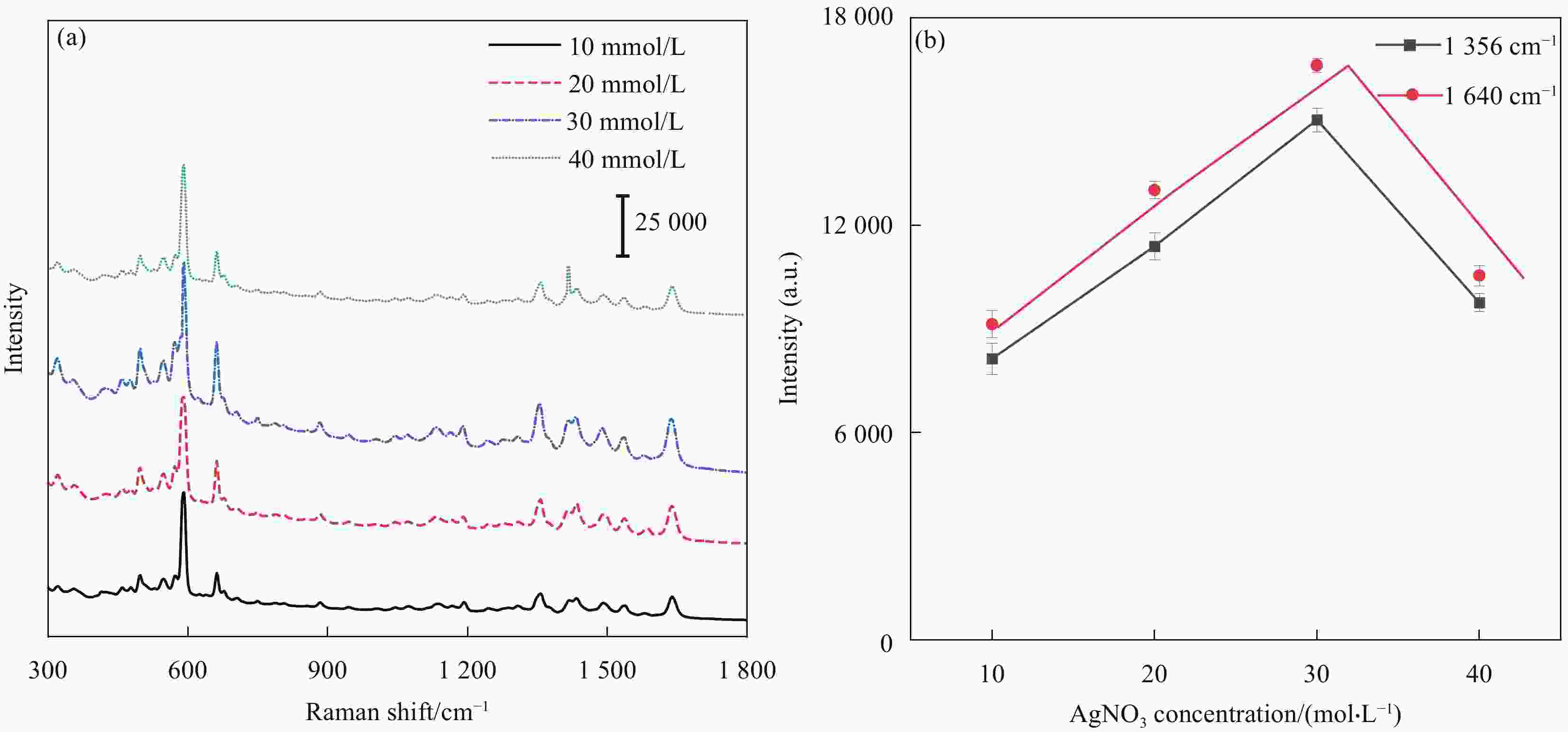
图 1 (a)不同浓度AgNO3制备的银/棉签复合材料的尼尔兰(NB)(10−6 mol/L)表面增强拉曼光谱(SERS) ; (b) 尼尔兰在1356, 1640 cm−1的拉曼强度与AgNO3浓度的关系图
Figure 1. (a) Surface enhanced Raman scattering (SERS) spectra of Nile Blue (NB) (10−6 mol/L) on Ag/cotton swab composite prepared with different concentrations of AgNO3; (b) Plots of Raman intensity of NB at 1356, 1640 cm−1 versus concentration of AgNO3
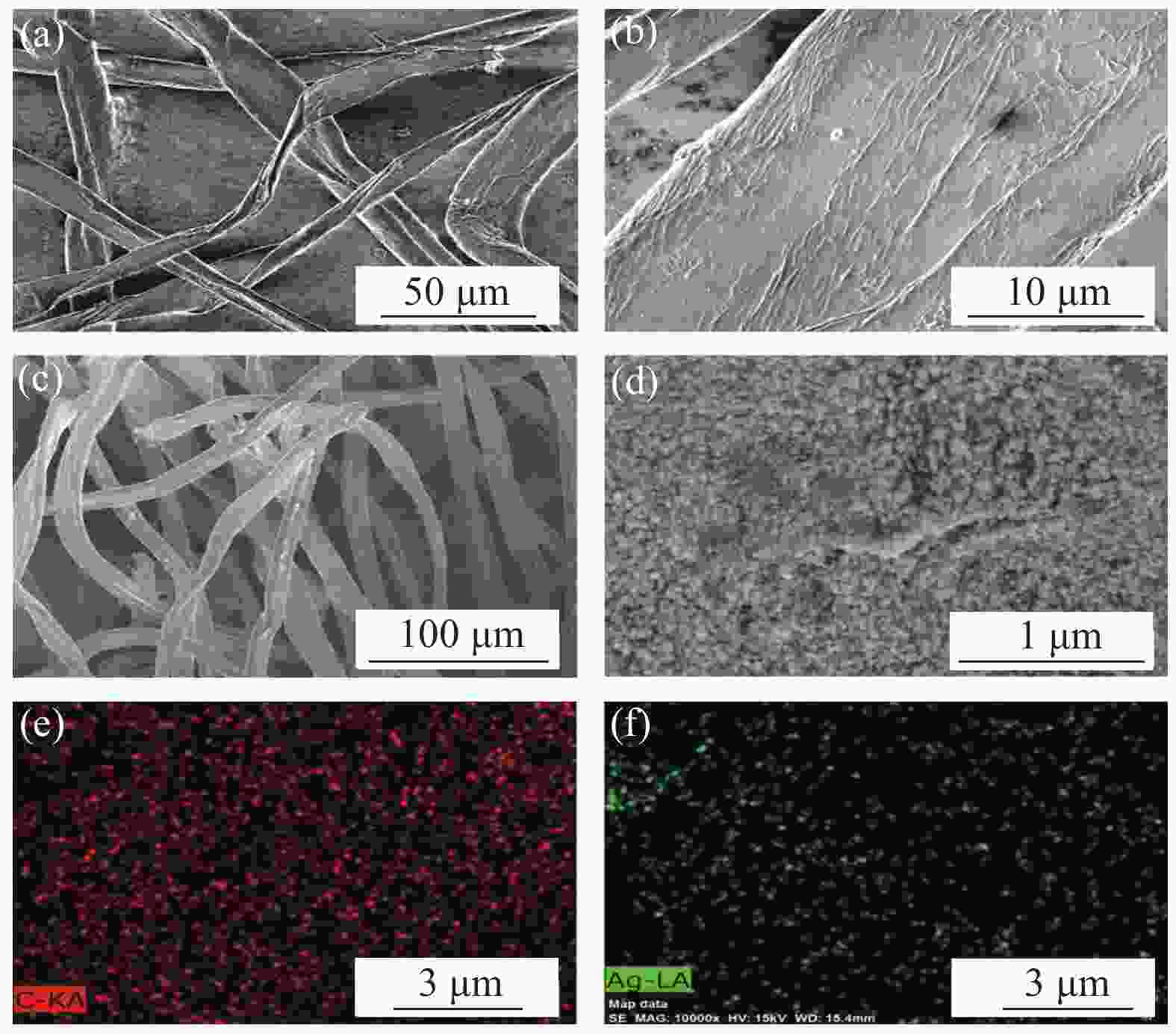
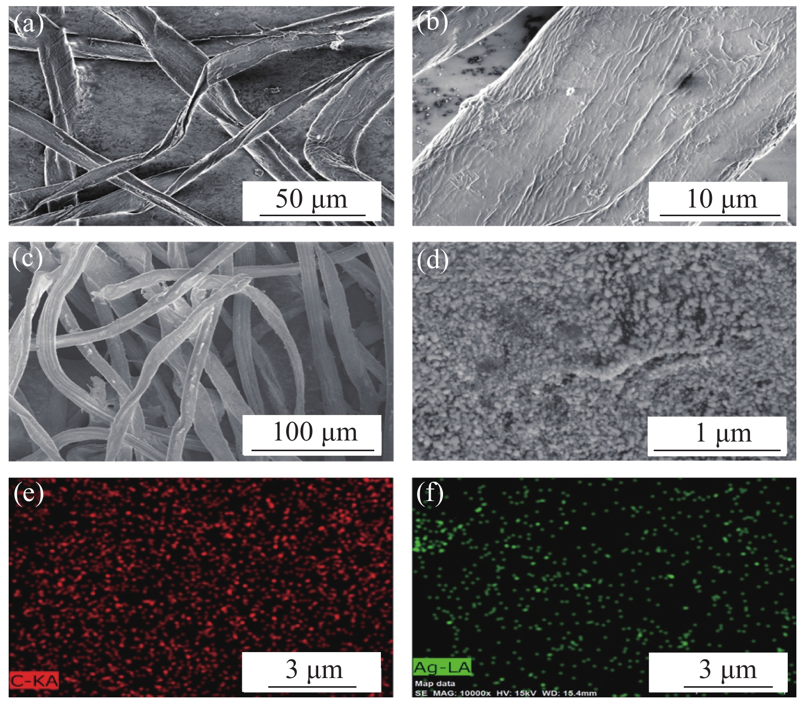
图 2 ((a), (b)) 棉签在沉积银纳米粒子前的SEM图像; ((c), (d)) Ag/棉签复合材料的SEM图像; ((e), (f)) Ag/棉签复合材料中Ag和C的元素成像
Figure 2. ((a), (b)) SEM images of the cotton swab before deposition of silver nanoparticles; ((c), (d)) SEM image of Ag/cotton swab composite; ((e), (f)) Elemental mapping of Ag and C in the Ag/cotton swab composite
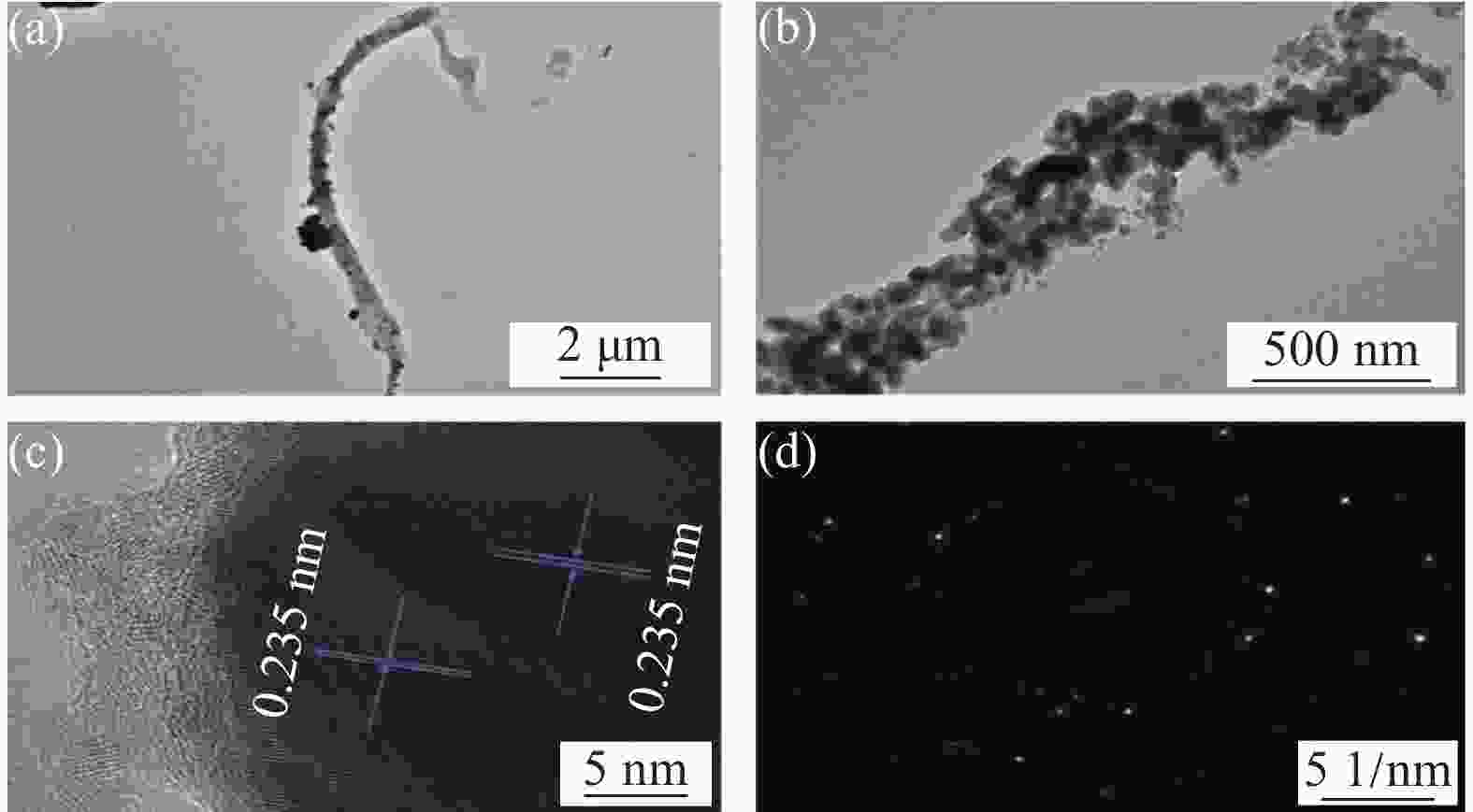
图 3 ((a), (b)) Ag/棉签复合材料的TEM图像; (c) 随机选择的银纳米粒子的HRTEM图像; (d) 棉签上银纳米粒子的选区电子衍射(SAED)图谱
Figure 3. ((a), (b)) TEM images of Ag/cotton swab composite; (c) HRTEM of randomly selected silver nanoparticle; (d) Selected area electron diffraction (SAED) spectrum of silver nanoparticle on cotton swab
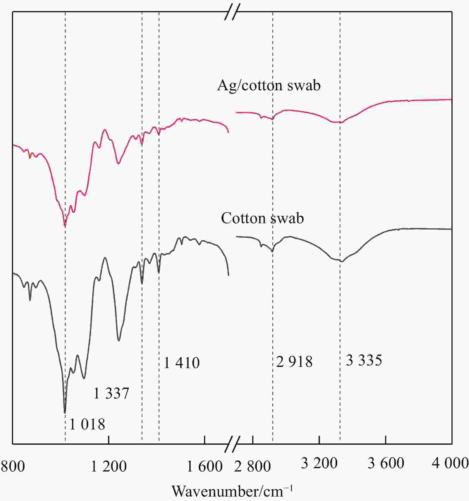
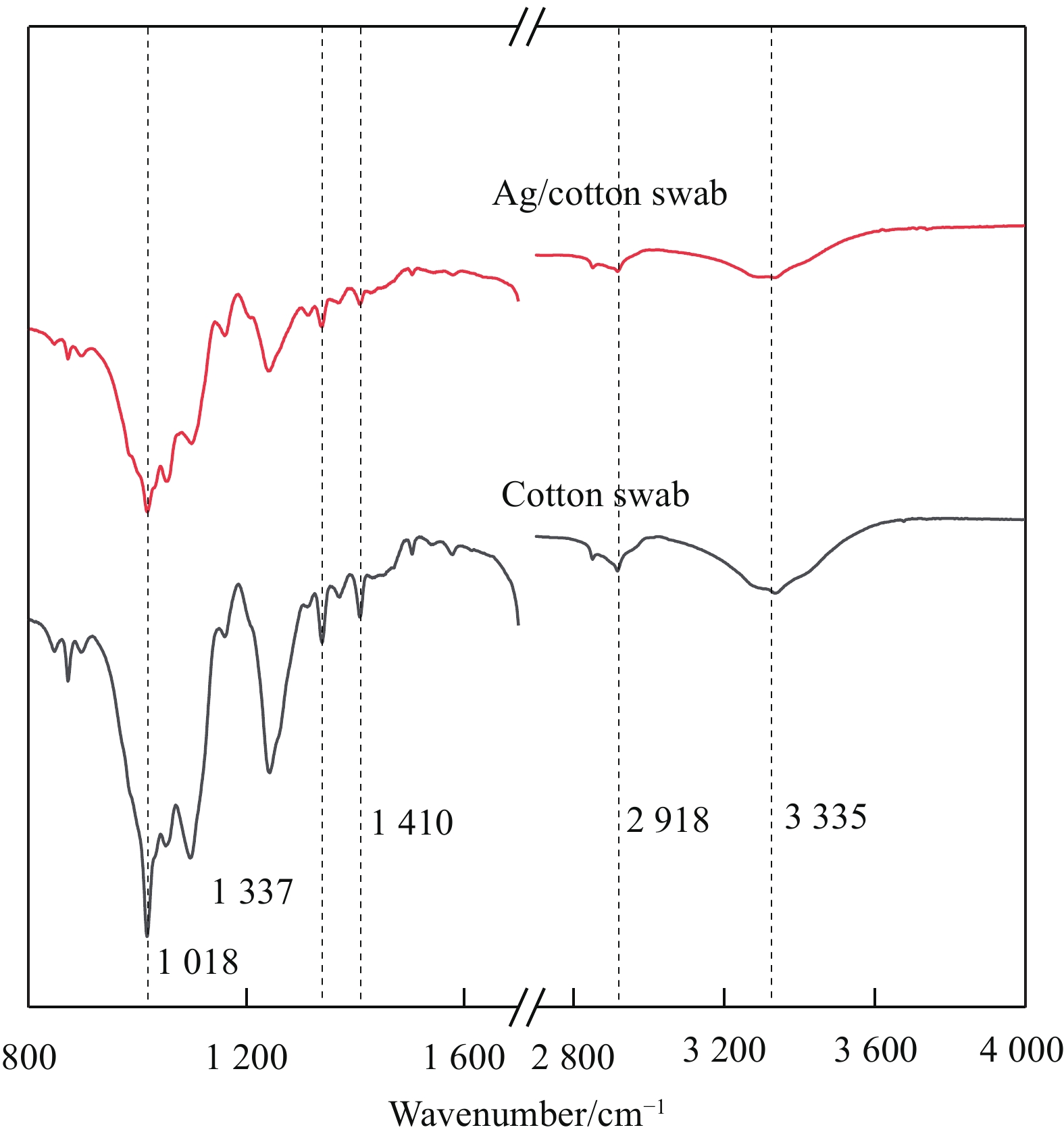
图 4 棉签和Ag/棉签复合材料的红外光谱
Figure 4. FT-IR spectra of cotton swab and Ag/cotton swab composite
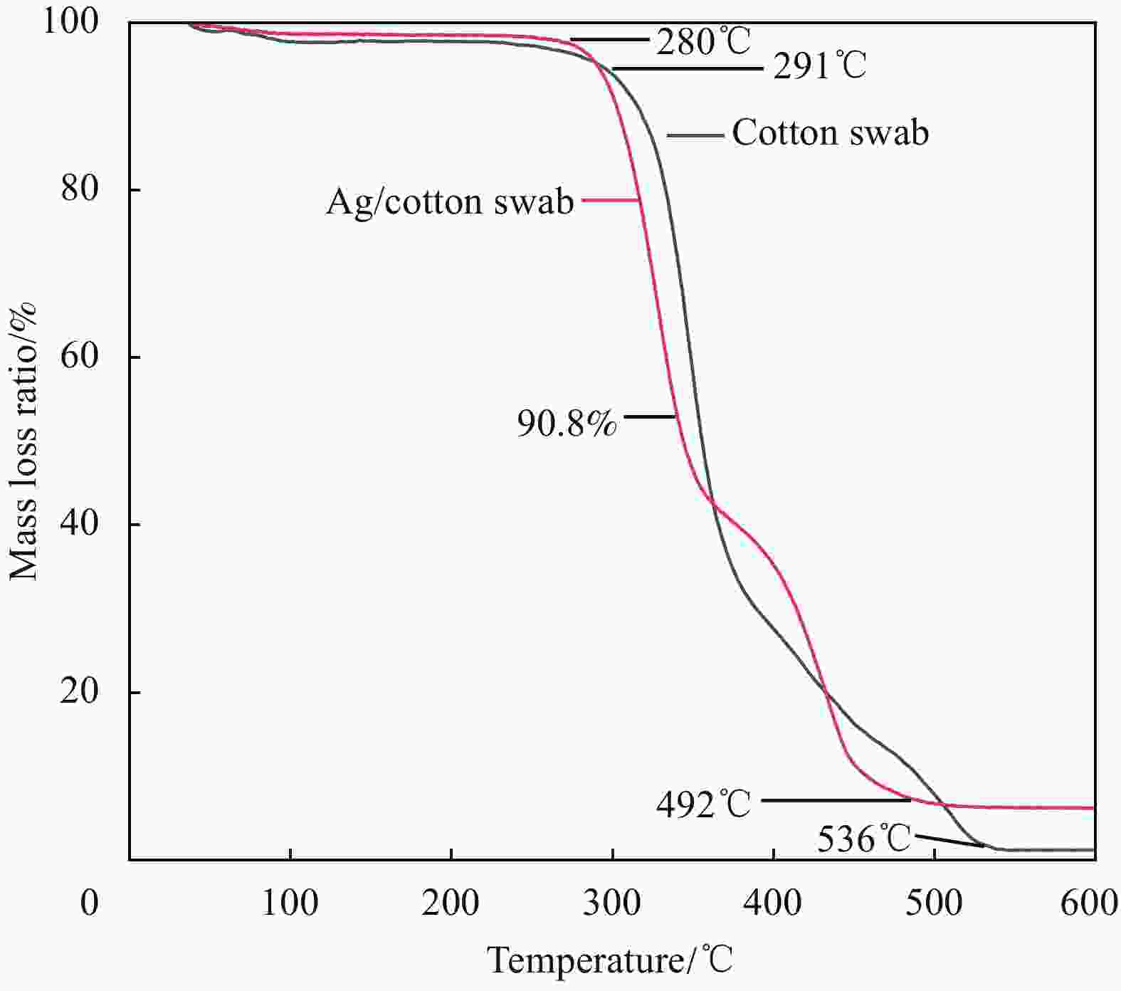
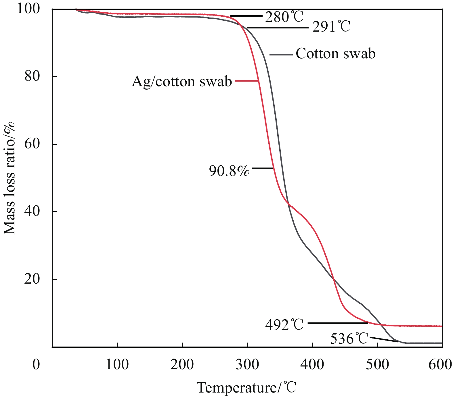
图 5 棉签和Ag/棉签复合材料的热重图(TG)
Figure 5. Thermogravimetry (TG) of cotton swab and Ag/cotton swab composite
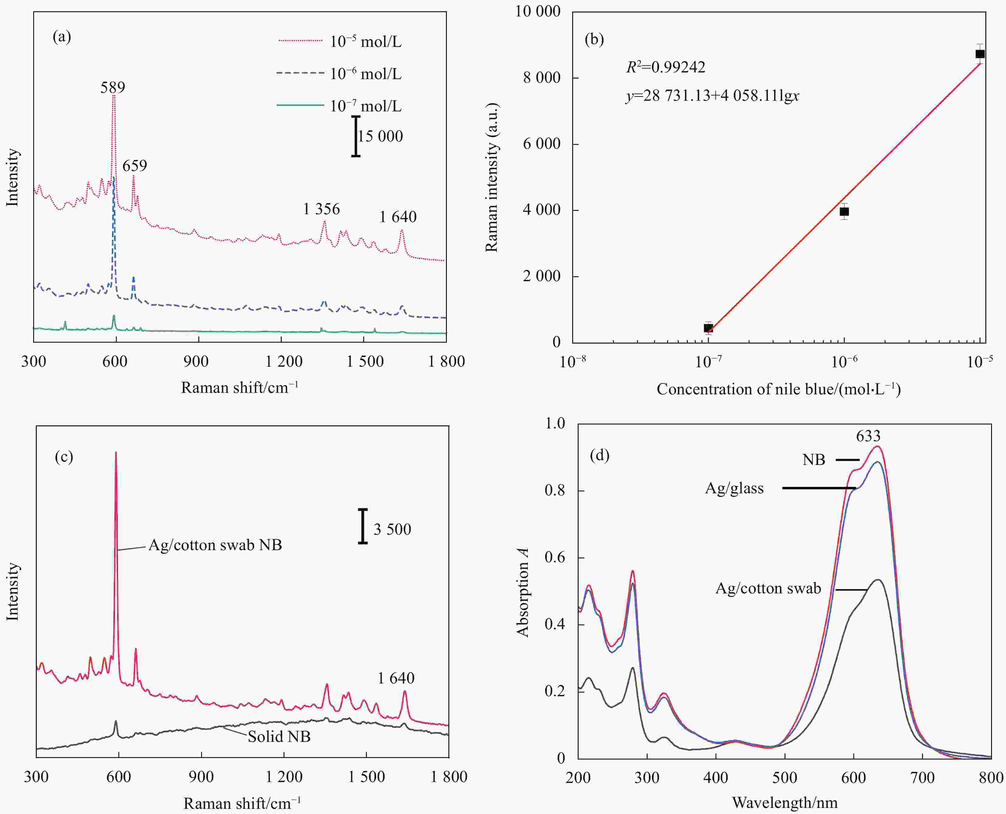
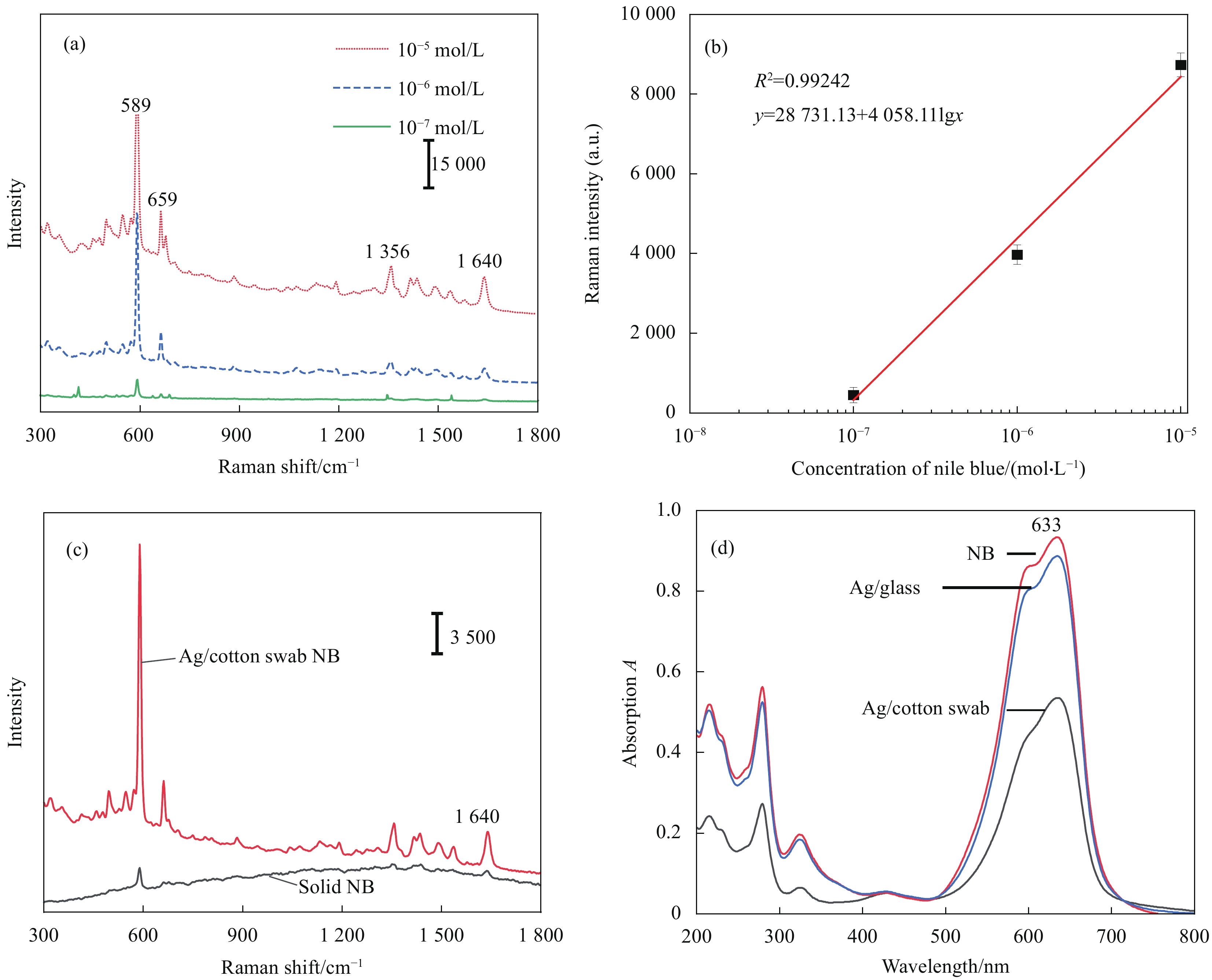
图 6 (a)不同浓度尼尔兰的SERS光谱; (b) 1640 cm−1处拉曼峰强度与尼尔兰浓度曲线; (c)固体尼尔兰和从Ag/棉签复合材料上测的尼尔兰的SERS光谱; (d)不同基底吸附后的NB(1.3×10⁻⁵ mol/L)吸收光谱
Figure 6. (a) SERS spectra of NB at different concentrations; (b) Dose-response curves of the peak at 1640 cm−1 from the spectra; (c) Solid NB and SERS spectra of NB measured from Ag/cotton swab composite; (d) Absorption spectra of NB (1.3×10⁻⁵ mol/L) after adsorbed by different substrate
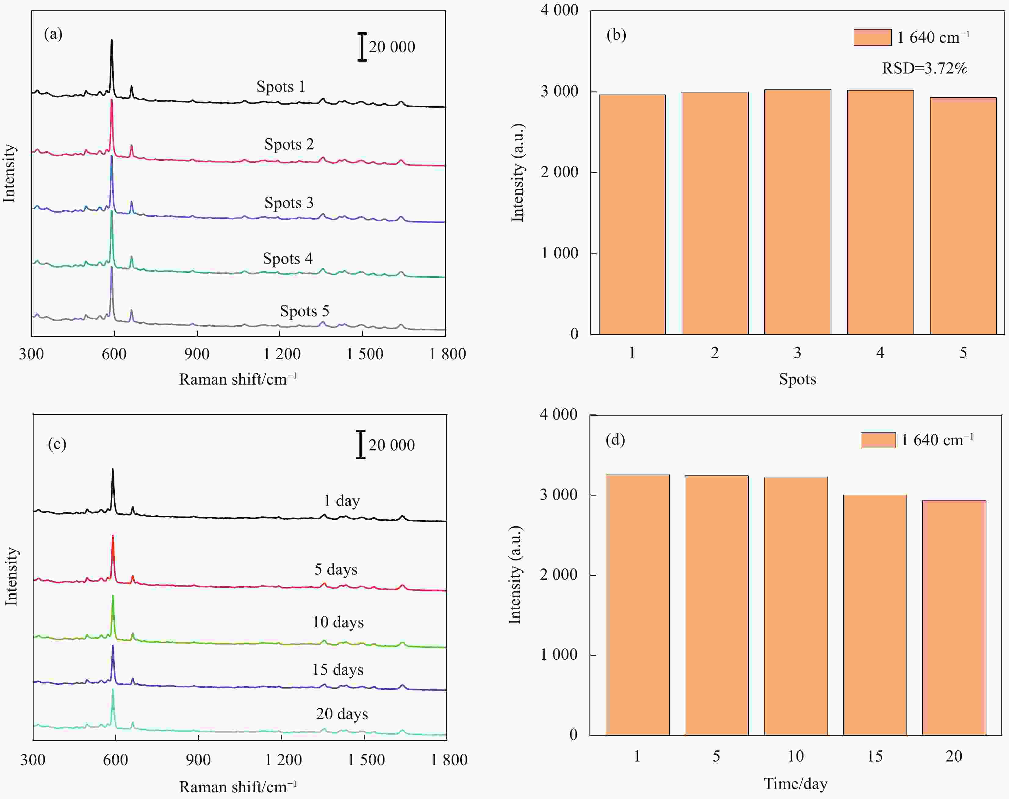
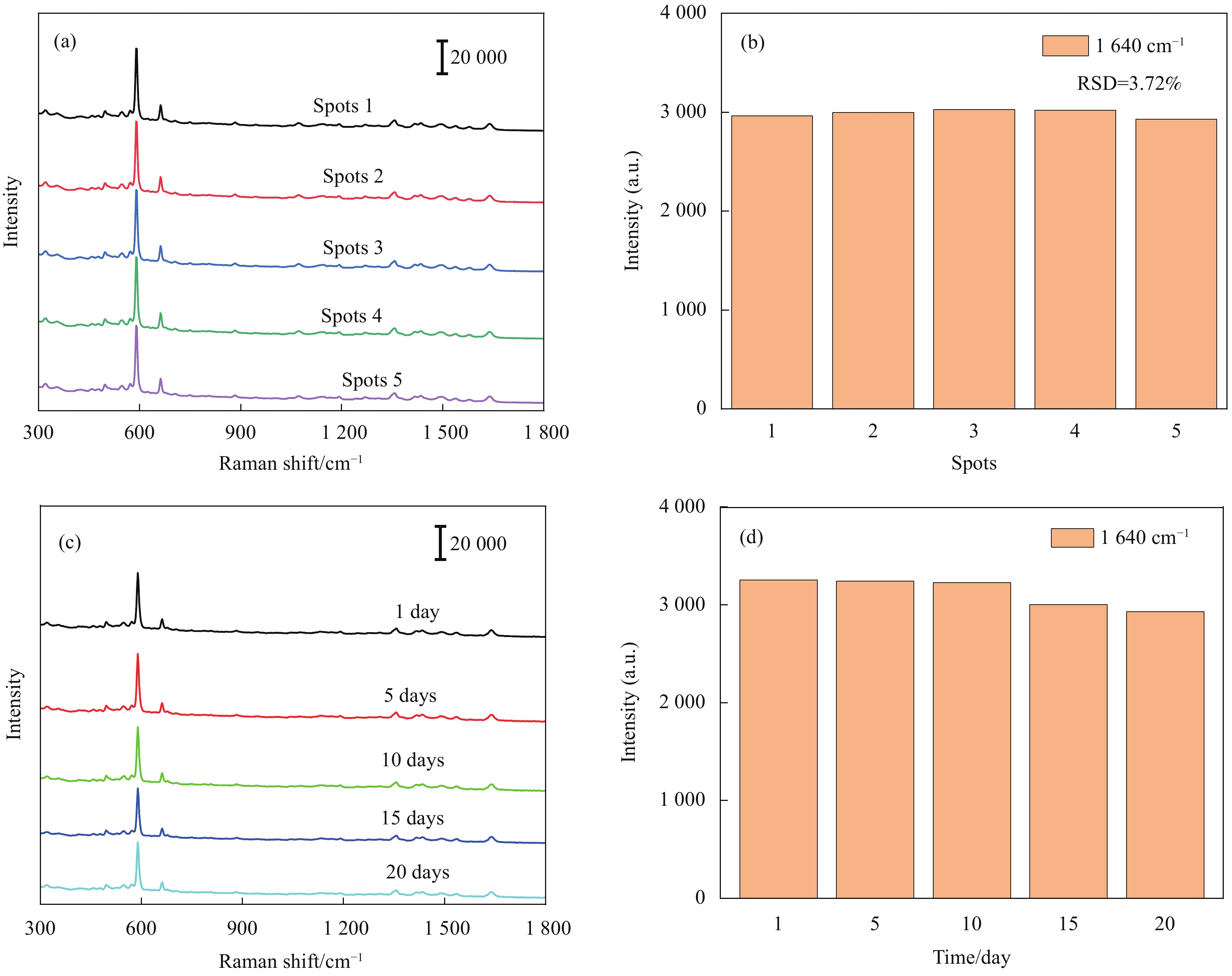
图 7 (a) Ag/棉签复合材料上五个随机选择位置尼尔兰(10−6 mol/L)的拉曼光谱; ((b), (d)) 强度统计图; (c) 20天内Ag/棉签复合材料上尼尔兰(10−6 mol/L)的拉曼光谱
Figure 7. (a) SERS spectrum of NB (10−6 mol/L) at five randomly selected positions on the Ag/cotton swab composite; ((b), (d)) Raman intensity statistics; (c) SERS spectra of NB (10−6 mol/L) during 20 days
RSD—Relative standard deviation
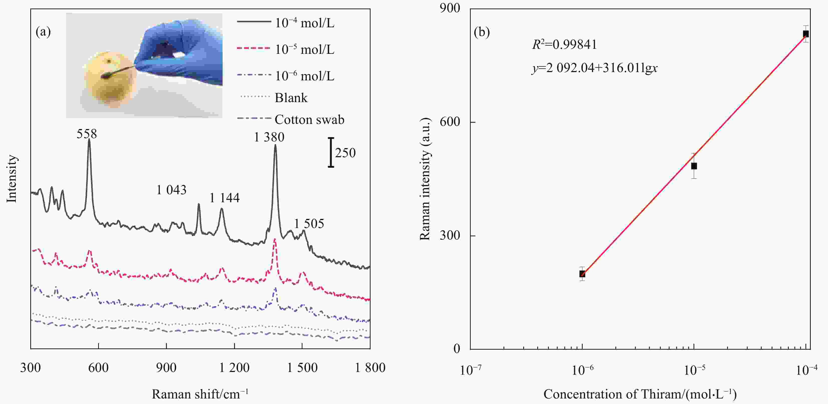
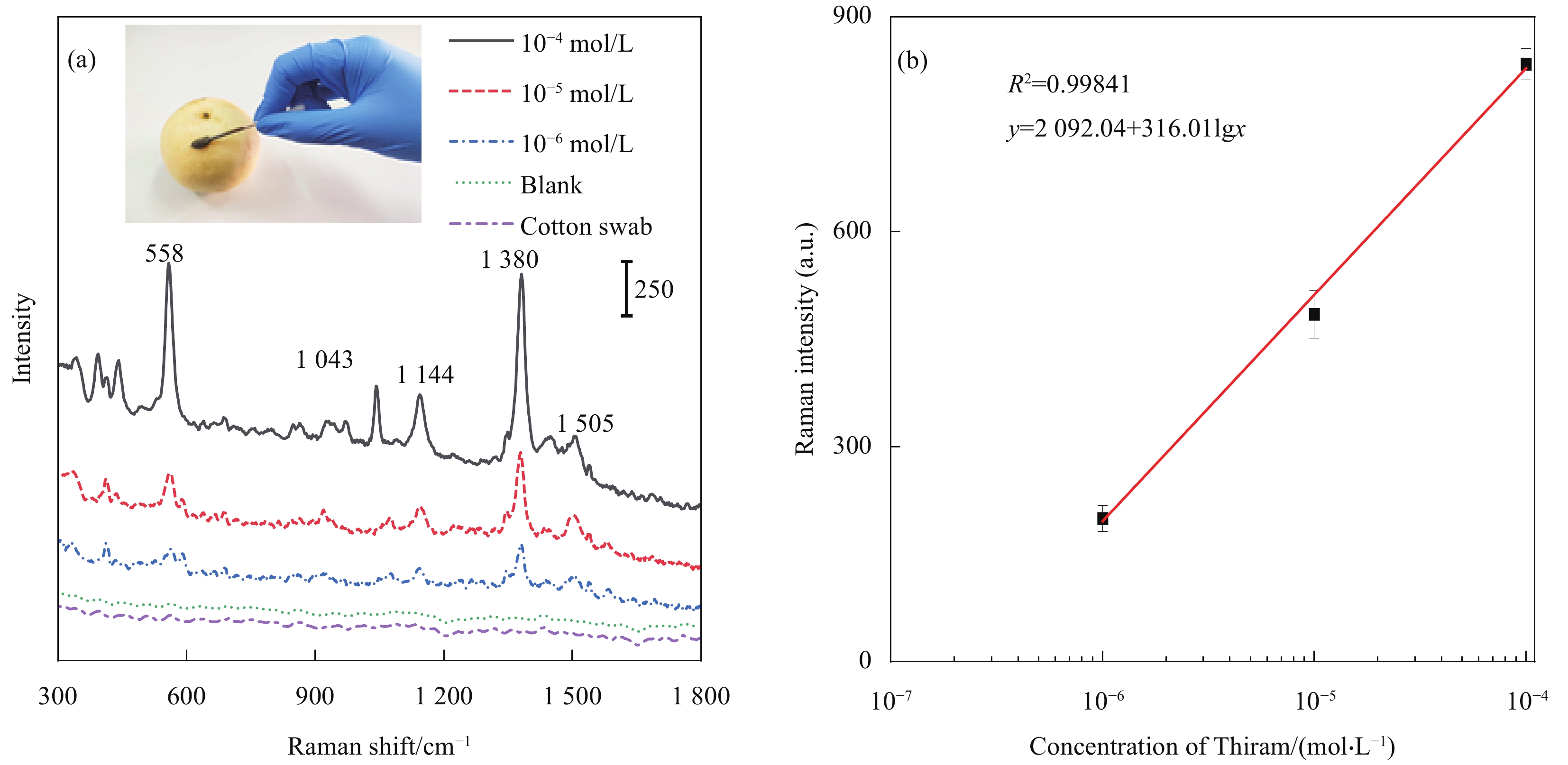
图 8 (a) Ag/棉签复合材料作为SERS基底从梨表面检测福美双; (b)梨表面测量的1380 cm−1处拉曼峰强度与福美双浓度曲线
Figure 8. (a) Ag/cotton swab composite used as SERS substrate to detect Thiram from the surface of pear; (b) Dose-response curves of the peak at 1380 cm−1 from the spectra measured from pear
-
[1] WERNE T V, PATTN T E. Preparation of structurally well-defined polymer-nanoparticle hybrids with controlled/living radical polymerizations[J]. Journal of the American Chemical Society,1999,121:7409-7410. doi: 10.1021/ja991108l [2] ZHANG J T, KELLER T F, GALLAGHER H, et al. Responsive hybrid polymeric/metallic nanoparticles for catalytic applications[J]. Macromolecular Materials and Engi-neering,2010,295(11):1049-1057. doi: 10.1002/mame.201000204 [3] WU J L, CHEN F C, HSIAO Y S, et al. Surface plasmonic effects of metallic nanoparticles on the performance of polymer bulk heterojunction solar cells[J]. ACS Nano,2011,5(2):959-967. doi: 10.1021/nn102295p [4] HARTMAN C, POPOWSKI Y, RAICHMAN D, et al. Biodegradable polymer coating for controlled release of hydrophobic functional molecules from cotton fabrics[J]. Journal of Coatings Technology and Research,2020,17:669-679. doi: 10.1007/s11998-019-00278-3 [5] ZHANG R, ZHANG Y, DONG Z C, et al. Chemical mapping of a single molecule by plasmon-enhanced Raman scattering[J]. Nature,2013,498:82-86. doi: 10.1038/nature12151 [6] GONG Z, DU H, CHENG F, et al. Fabrication of SERS swab for direct detection of trace explosives in fingerprints[J]. ACS Applied Materials & Interfaces,2014,6(24):21931-21937. doi: 10.1021/am507424v [7] FAN M, ZHANG Z, HU J, et al. Ag decorated sandpaper as flexible SERS substrate for direct swabbing sampling[J]. Materials Letters,2014,133(15):57-59. [8] LANE L A, QIAN X, NIE S. SERS nanoparticles in medicine: From label-free detection to spectroscopic tagging[J]. Chemical Reviews,2015,115(19):10489-10529. doi: 10.1021/acs.chemrev.5b00265 [9] ZHANG H, LIU M, ZHOU F, et al. Physical deposition improved SERS stability of morphology controlled periodic micro/nanostructured arrays based on colloidal templates[J]. Small,2015,11(7):844-853. [10] SUN F, HUNG H C, SINCLAI R, et al. Hier-archical zwitterionic modification of a SERS substrate enables real-time drug monitoring in blood plasma[J]. Nature Communications,2016,7:13437. doi: 10.1038/ncomms13437 [11] STRANAHAN S M, WILLETS K A. Super-resolution optical imaging of single-molecule SERS hot spots[J]. Nano Letters,2010,10(9):3777-3784. doi: 10.1021/nl102559d [12] JI W, LI L, SONG W, et al. Enhanced Raman scattering by ZnO superstructures: Synergistic effect of charge transfer and Mie resonances[J]. Angewandte Chemie Interna-tional Edition,2019,58(41):14452-14456. doi: 10.1002/anie.201907283 [13] JIANG X, ZHANG J, XU L, et al. Ultrasensitive SERS detection of antitumor drug methotrexate based on modified Ag substrate[J]. Spectrochimica Acta Part A: Molecular and Biomolecular Spectroscopy,2020,240:118589. doi: 10.1016/j.saa.2020.118589 [14] LIN X, FANG G, LIU Y, et al. Marangoni effect-driven transfer and compression at three-phase interfaces for highly reproducible nanoparticle monolayers[J]. Journal of Phy-sical Chemistry Letters,2020,11(9):3573-3581. doi: 10.1021/acs.jpclett.0c01116 [15] ZHANG C, YOU T, YANG N, et al. Hydrophobic paper-based SERS platform for direct-droplet quantitative determination of melamine[J]. Food Chemistry,2019,287:363-368. doi: 10.1016/j.foodchem.2019.02.094 [16] XIONG Z, LIN M, LIN H, et al. Facile synthesis of cellulose nanofiber nanocomposite as a SERS substrate for detection of thiram in juice[J]. Carbohydrate Polymers,2018,189:79-86. doi: 10.1016/j.carbpol.2018.02.014 [17] CHEN Y, GE F, GUANG S, et al. Self-assembly of Ag nanoparticles on the woven cotton fabrics as mechanical fle-xible substrates for surface enhanced Raman scattering[J]. Journal of Alloys and Compounds,2017,726:484-489. doi: 10.1016/j.jallcom.2017.07.315 [18] XIA L, XU M, CHENG G, et al. Facile construction of Ag nanoparticles encapsulated into carbon nanotubes with robust antibacterial activity[J]. Carbon,2018,130:775-781. doi: 10.1016/j.carbon.2018.01.073 [19] DUCHEMIN B, CORRE D L E, LERAY N, et al. All-cellulose composites based on microfibrillated cellulose and filter paper via a NaOH-urea solvent system[J]. Cellulose,2016,23:593-609. doi: 10.1007/s10570-015-0835-4 [20] CHENG D, HE M, RAN J, et al. Depositing a flexible substrate of triangular silver nanoplates onto cotton fabrics for sensitive SERS detection[J]. Sensors and Actuators B: Chemical,2018,270:508-517. doi: 10.1016/j.snb.2018.05.075 [21] ZHOU Y, ZHI J, ZHAO J, et al. Surface-enhanced Raman scattering of 4-aminothiophenol adsorbed on silver nanosheets deposited onto cubic boron nitride films[J]. Analytical Sciences,2010,26(9):957-961. doi: 10.2116/analsci.26.957 [22] 刘思佳, 喻倩, 王锐, 等. 再生纤维素纤维-纳米金柔性复合物的制备及其对尼尔兰的快速检测[J]. 纺织学报, 2020, 41(7):24-28.LIU Sijia, YU Qian, WANG Rui, et al. Preparation of flexible Au nanoparticle decorated regenerated cellulose fiber compound and quickly detection of Nile Blue[J]. Journal of Textile Research,2020,41(7):24-28(in Chinese). [23] LI D, OUYANG L, YAO L, et al. In situ SERS monitoring the visible light photocatalytic degradation of Nile Blue on Ag@AgCl single hollow cube as a microreactor[J]. Chemistry Select,2018,3(2):428-435. doi: 10.1002/slct.201702545 [24] ZHU Y, LI M, YU D, et al. A novel paper rag as ‘D-SERS’ substrate for detection of pesticide residues at various peels[J]. Talanta,2014,128:117-124. doi: 10.1016/j.talanta.2014.04.066 [25] SUN H, LIU H, WU Y. A green, reusable SERS film with high sensitivity for in-situ detection of thiram in apple juice[J]. Applied Surface Science,2017,416:704-709. doi: 10.1016/j.apsusc.2017.04.159 -

 下载:
下载:


 点击查看大图

计量
- 文章访问数: 663
- HTML全文浏览量: 468
- PDF下载量: 31
- 被引次数: 0
